Palliative care, also known as supportive care, helps people with serious illnesses feel more comfortable. Palliative care can help relieve pain and treat side effects as people go through disease treatment or hospice care at the end of life. Palliative dental care can be crucial for patients fighting cancer or other diseases because they can have unique and troublesome dental issues. Read more about palliative dental care so you can prepare for a loved one who may need it someday.
The Role of Supportive Care in Dentistry
Unfortunately, people receiving treatment for cancer or other serious illnesses may experience unpleasant or painful side effects. Dentists and dental hygienists treat these side effects to relieve this discomfort and anxiety during an already stressful time. Palliative care physicians can refer a family to a dentist, or the patient's dentist may create a new care plan that supplements other palliative care treatments.
When Palliative Dental Care May Help
According to the Journal of the American Geriatrics Society, many oral health problems can affect patients with serious illnesses. These are the typical side effects during cancer treatment that palliative dentistry can help with: Dry mouth, Mouth sores or inflammation, Candidiasis or oral thrush, Severe tooth decay.
According to The Oral Cancer Foundation (OCF), constant dry mouth is a common side effect of radiation therapies. More than 400 medications may cause dry mouth and up to 40 percent of patients undergoing chemotherapy report mouth sores and inflammation. This side effect is called mucositis, and it can interfere with eating, swallowing, and talking comfortably.
Cancer patients or people with immune disorders may develop candidiasis, also known as oral thrush. Oral thrush can make eating and drinking uncomfortable because of itching and burning sensations.
Lastly, people with severe or terminal illnesses may be more susceptible to tooth decay. For example, patients undergoing radiation therapy can develop what is called radiation caries. If not treated quickly, these cavities can lead to tooth abscesses. These side effects and dental issues may need special or more frequent treatment from a dentist and dental hygienist.
Supportive Dental Care Procedures
Palliative treatment, including dental care, focuses on managing the symptoms of these side effects and issues to keep palliative patients comfortable. The dentist can recommend a particular dental care plan after examining and talking with you or your loved one's healthcare team. Some of the palliative dental care treatments can include: More frequent checkups and preventive care, Recommendations of mouthrinse for dry mouth, Antibiotics for mouth sores, Anti-fungal medications for oral thrush.
Along with these treatments, the dentist may suggest drinking more water or eating a bland, soft diet to relieve dry mouth and relieve discomfort from mouth sores.
Dental restorations such as fillings or crowns may be required for patients who have developed cavities. In cases of advanced decay where an abscess has formed, the dentist may remove the tooth.
Palliative dental care can help relieve the pain and discomfort from the side effects of cancer treatments and other ailments. With palliative care, patients can typically eat, drink, and talk more comfortably. If your loved one is considering palliative care with hospice or with their doctors, speak to their dentist about maintaining their oral health too.
Your body is a complex system with various components working together to keep it performing at its optimum best. The parathyroid hormone (PTH) helps maintain calcium and phosphorus metabolism. It has an important role in bone and teeth mineralization. But what happens if the parathyroid gland isn't working properly? Various conditions point to problems with the PTH, like hypercalcemia. Learn more about hypercalcemia, its effects on your oral health, and treatment options.
What Is Hypercalcemia?
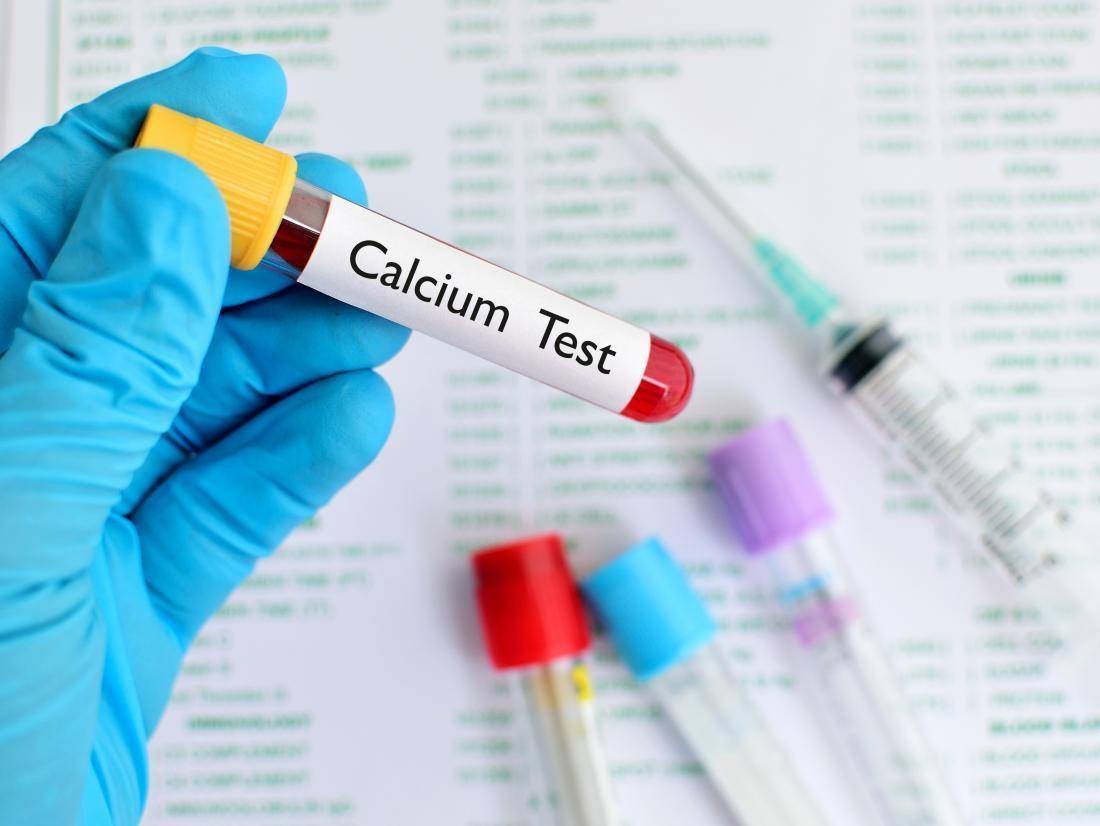
Hypercalcemia is a condition marked by increased levels of calcium in the blood. The parathyroid gland regulates the calcium levels in your body by releasing parathyroid hormones (PTH). Issues with the parathyroid gland can cause an excessive release of the parathyroid hormone, also known as hyperparathyroidism (HPT). There are three types of hyperparathyroidism: primary, secondary, and tertiary. Primary hyperparathyroidism (PHPT) and cancer are the most common causes of hypercalcemia, accounting for over 80-90% of cases.
Hypercalcemia and Teeth
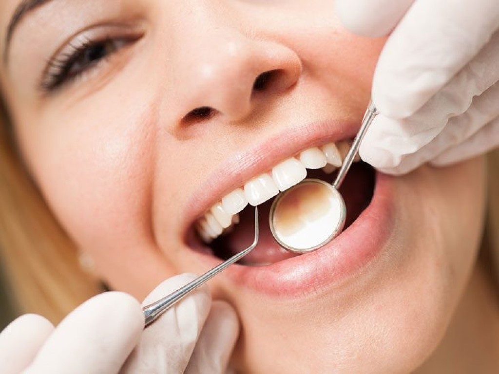
How does hypercalcemia affect your oral health? Calcium is one of the things your body needs to build strong, healthy teeth and bones. It also builds and maintains your jawbone, helping to create a solid anchor for your teeth. So what happens if you have too much calcium in the teeth? There are mild to severe oral symptoms that your dentist is specially trained to detect and treat. People with varying types of hyperparathyroidism and hypercalcemia may experience:
Soft tissue calcifications
Tooth sensitivity when biting and chewing
Malocclusion
Cavities
Slight jaw pain
Treatment for Hypercalcemia
Your doctor will recommend a treatment option based on the severity and cause of the condition. According to the Cleveland Clinic, your doctor may suggest the following to moderate your calcium levels
Drink more water: This will help flush out the excess calcium.
Avoid calcium supplements.
Avoid calcium-rich antacid tablets.
To ensure that your overall health is taken care of, your doctor will monitor and treat the underlying causes of hypercalcemia too.
A hormonal imbalance and some cancer and cancer treatments may lead to hypercalcemia. Fortunately, your dentist can detect and effectively treat oral issues due to these conditions. Talk to your doctor and dentist if you are experiencing discomfort in your teeth when chewing, pain in the jaw, or any other dental problems. They can help you take good care of your oral health while undergoing treatment.
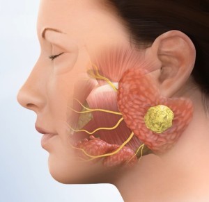
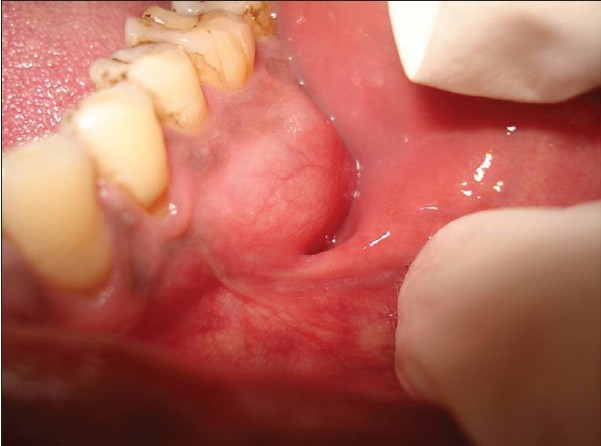
Parotid tumors are abnormal growths of cells (tumors) that form in the parotid glands. The parotid glands are two salivary glands that sit just in front of the ears on each side of the face. Salivary glands produce saliva to aid in chewing and digesting food.
There are many salivary glands in the lips, cheeks, mouth and throat. Tumors can occur in any of these glands, but the parotid glands are the most common location for salivary gland tumors. Most parotid tumors are noncancerous (benign), though some tumors can become cancerous.
Parotid tumors often cause swelling in the face or jaw that usually isn't painful. Other symptoms include numbness, burning or prickling sensations in the face, or a loss of facial movement.
Diagnosis
Tests and procedures used to diagnose a parotid tumor may include:
A physical exam. Your doctor will feel your jaw, neck and throat for lumps or swelling.
Collecting a sample of tissue for testing (biopsy). Your doctor may recommend a needle biopsy procedure, such as fine-needle aspiration or core needle biopsy, to collect a sample of tissue for testing. During a needle biopsy, the doctor inserts a thin needle through your skin and into the affected parotid gland. The needle is used to draw out a sample of cells or fluid.
In the lab, doctors can determine what types of cells are involved and whether they're cancerous. Your doctor uses this information to determine your prognosis and which treatments are best for you.
Imaging tests. Your doctor may recommend imaging tests of your parotid gland to help understand the size of your tumor. If your parotid tumor is cancerous, you may need tests to look for signs that the cancer has spread. Tests may include ultrasound, MRI and CT.
Treatment
Parotid tumor treatment usually involves surgery to remove the tumor. If the tumor contains cancer cells, your doctor may recommend additional treatments, such as radiation therapy and chemotherapy.
Surgery
Operations used to remove parotid tumors include:
Removing part of the parotid gland. For most parotid tumors, surgeons may cut away the tumor and some of the healthy parotid gland tissue around it (superficial parotidectomy).
Removing all of the parotid gland. Surgery to remove all of the parotid gland (total parotidectomy) might be recommended for larger tumors and those that affect the deeper parts of the parotid gland.
More extensive surgery for larger cancers. If parotid cancer has grown into nearby bone and muscles, a more extensive operation may be necessary. Surgeons try to remove all of the cancer and a small amount of the healthy tissue that surrounds it. Then they work to repair the area so you can continue to chew, swallow, speak, breathe and move your face. This may involve transferring skin, tissue, bone or nerves from other parts of your body to make repairs.
To access the parotid gland, surgeons make an incision near the ear. During the operation, special care is taken to avoid damage to nearby structures, such as the facial nerve that runs through the parotid gland. The facial nerve controls facial movement, so stretching or cutting the nerve can cause partial or complete paralysis of the face that can be temporary or permanent.
If the facial nerve must be cut in order to remove all of the tumor, surgeons can repair it using nerves from other areas of your body or processed nerve grafts from donors.
Radiation therapy
Radiation therapy uses powerful beams of energy, such as X-rays, protons or neutrons, to kill cancer cells. If your parotid tumor is cancerous, radiation therapy might be recommended after surgery to kill any cancer cells that remain. Radiation therapy is sometimes used as an initial treatment when surgery isn't an option.
Chemotherapy
Chemotherapy is a drug treatment that uses medications to kill cancer cells. It's not routinely used to treat parotid tumors. But sometimes it's combined with radiation therapy to treat parotid cancers that have a high risk of spreading or cancers that can't be removed completely with surgery. Chemotherapy might also be an option for people with advanced parotid cancers that have spread to other parts of the body.
The rarity of verrucous carcinoma. It's not just limited to tobacco-chewing ballplayers, but anyone prone to smoking cigarettes or chewing tobacco. Learn the symptoms and how you can strike it out if it comes to your plate.
What Is Verrucous Carcinoma?
Verrucous carcinoma is an uncommon cancer that often develops in an area of extreme irritation or inflammation with symptoms of cauliflower-like lesions. It's so rare that the American Cancer Society says it accounts for less than 5% of oral cancers. The most typical place for it to appear is within the oral cavity — or the larynx, nasal cavity, and throat. Patients with ill-fitting dentures, oral ulcerative areas, chronic candidiasis, and those who regularly smoke, chew tobacco, and consume alcohol are prone to develop verrucous carcinomas.
How Is Verrucous Carcinoma Treated?
The way to navigate the biggest threat to treating verrucous carcinomas is to avoid chewing and smoking tobacco. That's easier said than done — but necessary. The entire lineup of treatments to face verrucous carcinomas, though, is a powerful one.
Treatments include:
Surgery to remove the tumorous legions
Radiation therapy
A proper diet
Sufficient rest
What Should You Expect With Verrucous Carcinoma?
According to the American Cancer Society, verrucous carcinoma can easily be picked off. This means it's pretty slow-moving and rarely spreads throughout your body. That's good news. The bad news — it can grow deep within your oral tissue. A proper oral cancer screening with your dentist will help determine if you've developed any of these cancerous lesions. If you have developed verrucous carcinoma, you should seek treatment and immediately remove the lesions and surrounding normal tissue.
Proper brushing and flossing, as well as regular dental checkups, can only help you become an all-star in oral hygiene. But the most important thing you can do for your oral and overall health is to quit smoking and chewing tobacco immediately. Those two, along with a robust treatment plan — that's three strikes against verrucous carcinoma. It's out.
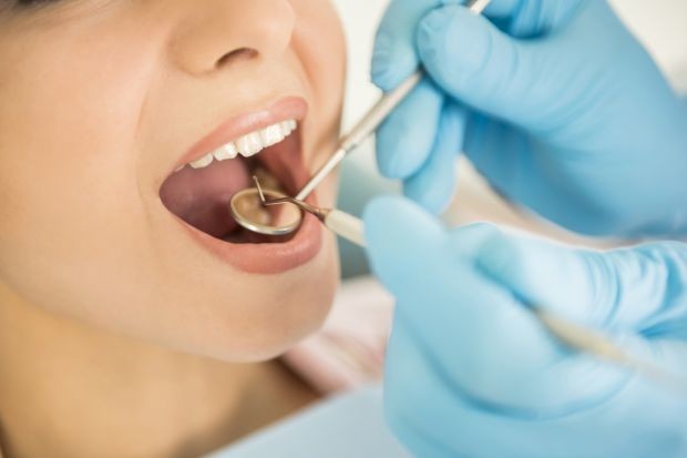
Each year, about 53,000 Americans are diagnosed with oral or oropharyngeal cancer, according to the Oral Cancer Foundation. Your dentist or doctor may find these cancers during a routine exam. Often, symptoms such as lingering mouth sores or red and white patches on the tongue, lips, or gums are the first indication something is wrong.
To determine whether these abnormalities are precancerous, cancerous, or benign, you’ll need to have an oral biopsy.
What Is Oral Biopsy?
Oral cancers are a type of head and neck cancer. They include cancer in your mouth and cancer in your oropharynx — the part of the throat that includes the tonsils, base of the tongue, and the soft palate.
If cancer is suspected, your dentist or doctor will refer you to a specialist. You will be referred to either an oral and maxillofacial surgeon or an otolaryngologist — an ear, nose, and throat (ENT) doctor. Both types of specialists are trained in medicine and surgery of the head and neck. The specialist will first conduct a thorough head and neck exam.
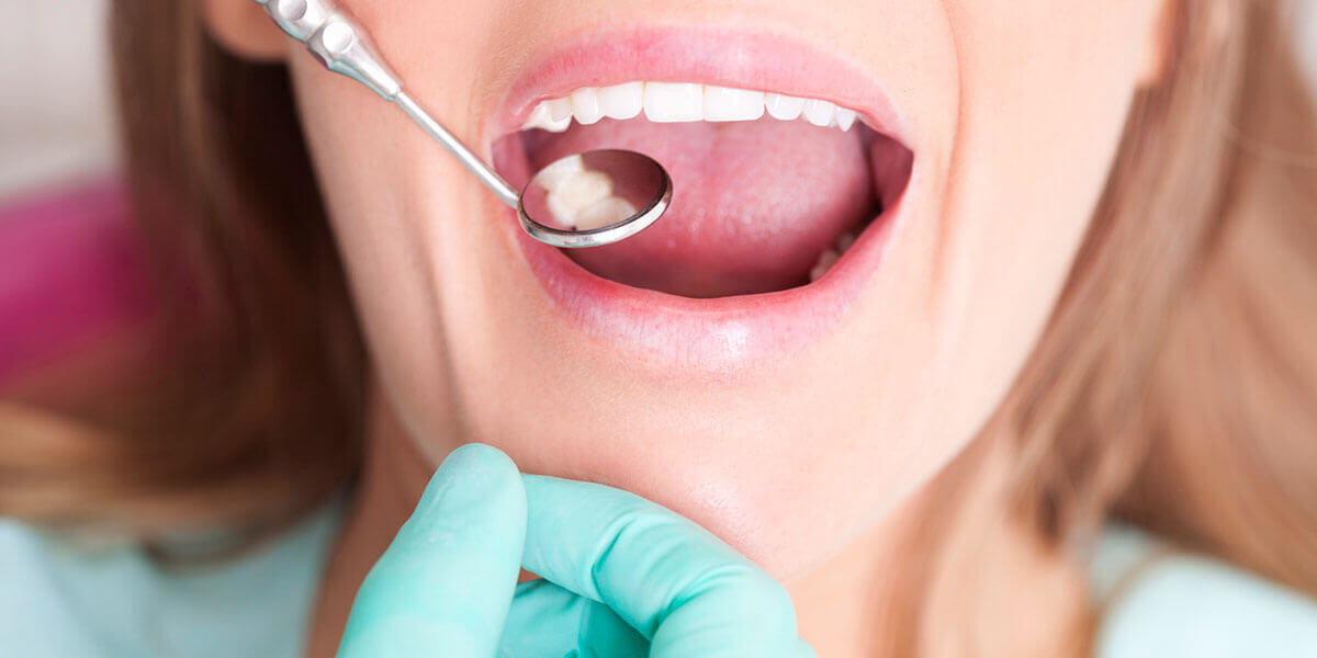
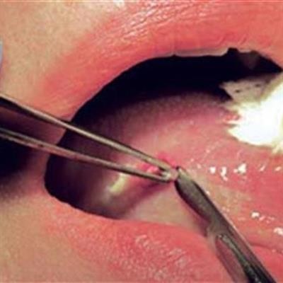
During an oral biopsy, the specialist removes a small amount of the suspicious tissue from your mouth or oropharynx and sends it to a pathologist, who will check for cancer cells. If cancer is confirmed, information in the pathologist’s report will help determine treatment.
Types of Oral Biopsies
There are three types of biopsies, according to the American Cancer Society. All are performed as outpatient procedures:
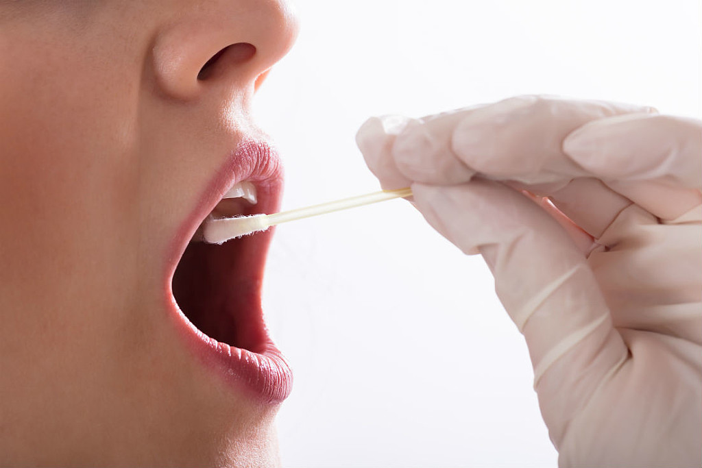
Exfoliative cytology: During this in-office procedure, the doctor gently scrapes cells from the suspicious area. It’s a quick, noninvasive, and painless way to check for oral cancer, especially if the lesion or area looks only slightly suspicious. Because this method doesn’t detect all oral cancers, you may need to undergo a more invasive biopsy.
Incisional biopsy: For this procedure, the doctor cuts out a sample of tissue for testing. Depending on the location of the suspicious tissue, an incisional biopsy can be performed either in a doctor’s office using local anesthesia or under general anesthesia in an operating room. In some cases, your doctor may have to perform an excisional biopsy, which requires cutting out most of the suspected area as well as some surrounding healthy tissue. This procedure may require stitches, which usually dissolve on their own within 10 days.
Fine-needle aspiration (FNA) biopsy: If you have a lump in your neck, your doctor may order an FNA biopsy. During this in-office procedure, the doctor will use a very thin needle to draw out fluid or cells from the lump. The doctor numbs the affected area beforehand.
In-office oral biopsies typically don’t require any preparation. Before any biopsy, your doctor will review your health records and tell you medicines to withhold. If you’ll be having general anesthesia, the doctor also will tell you how long to fast before the procedure.
Is an Oral Biopsy Painful?
You shouldn’t feel pain during an oral biopsy. You may, however, feel a sharp pinch or pin prick from the needle used to inject the local anesthetic or the needle used to take the biopsy, according to the Radiological Society of North America. You also may feel some pressure from the instruments used to collect the sample.
Some people may experience pain after the anesthesia wears off, depending on where in the mouth or throat the sample was removed. The biopsy site may be sore for several days, which could make it difficult for you to eat solid foods. Tylenol is usually sufficient to manage the pain. Avoid taking NSAIDs, such as aspirin, Advil, or Aleve, which can increase the chance of bleeding.
Suppose you notice an abnormal growth in your mouth, and your physician tells you it's ameloblastoma. You might be wondering, what does that mean, and is it serious? Read on to learn more about this odontogenic tumor and how your doctor can help you get the treatment you need.
What Is Ameloblastoma?
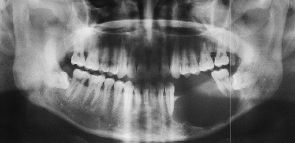
Ameloblastoma is a rare odontogenic tumor, which means it's formed from the normal tissues found in the mouth. Their location is predominately the lower jaw, also known as the mandible. Still, they can be found in the upper jaw or maxilla as well. Sometimes, ameloblastomas are located in the soft tissues surrounding the jaws. There is no known cause for this kind of tumor. However, it may be associated with impacted third molars. Ameloblastomas are usually noncancerous (benign), and they generally affect people between 40 and 60 years old.
Ameloblastoma Symptoms and Diagnosis
Ameloblastoma is usually painless, with the only symptom being swelling in the area. It is usually only identified on radiographic examination in a dental office. Early developing lesions do not displace teeth or cause numbness, so the patient may not know a tumor is growing in one of their jawbones. If a potential lesion is identified on a dental radiograph, more elaborate imaging is required. This will include a CAT scan and possibly an MRI. However, the diagnosis cannot be solely determined by imaging. It requires a biopsy to make the final diagnosis. Cysts will sometimes appear similar to the ameloblastoma on the imaging.
Some ameloblastomas are known to have malignant variants that are difficult to control locally. Metastases may occur, usually in the lung, but can spread through lymph nodes to other organs.
How Do You Treat Ameloblastoma?
Unfortunately, surgery is the only treatment to remove an ameloblastoma and prevent a recurrence. The procedure requires removing the healthy bone surrounding the tumor so that no tumor cells are left behind to allow it to regrow. Because the surgery must be performed aggressively, teeth will be extracted, and extensive hard and soft tissue plastic surgery reconstruction may be necessary. After the surgery, an opening into the upper jaw's sinuses requires the patient to wear a special denture known as an obturator. Unfortunately, recurrences are common even 10 to 20 years after surgery. Therefore, patients should be followed indefinitely and regularly evaluated for signs of changes to their mouth
To keep your oral and overall health in good shape, visit your dental professional regularly for oral health evaluations. Also, keep a watchful eye when you brush your teeth twice daily. Suppose you or your health care provider spot any abnormality. In that case, they can determine the cause and begin the necessary treatment to get you started on the road to recovery.
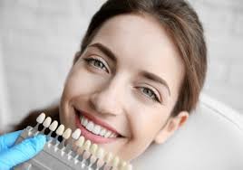
You know that you should see your dentist twice a year to get your teeth cleaned and checked out. Working with a general dentist can help you keep your smile as healthy as can be. But what can you do if you're concerned about the way your teeth look? Just as you'd see a cosmetic surgeon for aesthetic treatments on the body, you might see a cosmetic dentist for aesthetic treatments that improve the appearance of your teeth.
Learn more about the differences between a general dentist vs. cosmetic dentist, and find out if you need to see one or the other for the treatment you want.
What Do General and Cosmetic Dentists Do?
Your general dentist is your go-to resource for your overall oral health. As the British Columbia Dental Association notes, a general dentist typically diagnoses oral diseases, provides preventive treatments (such as cleanings), examines and interprets your X-rays and puts together treatment plans to help you achieve or maintain your oral health.
A cosmetic dentist focuses on the appearance of your teeth and gums. As the American Academy of Cosmetic Dentistry (AACD) describes, cosmetic dentists provide a wide range of aesthetic procedures, such as teeth whitening, dental implants, teeth bonding and veneers.
Educational Requirements for a General Dentist
To become a dentist, a person needs to graduate from an accredited dental school and earn either a Doctor of Dental Surgery (DDS) degree or a Doctor of Medicine in Dentistry (DMD) degree. As the American Dental Association (ADA) points out, DDS and DMD degrees are the same thing. Schools simply select which term to use for the degree.
Earning a dental degree prepares a person for a career as a general dentist. Just as some medical doctors pursue additional training to specialize in a particular area, some dentists may also choose to specialize in certain treatment areas. For example, they can complete additional training so that they can treat gum disease, provide orthodontic treatment or replace missing teeth. The ADA notes that there are 10 recognized dental specialties. Depending on the subject, a dentist can complete anywhere from one to eight years of additional education to earn certification in a specialty, as the ADA outlines.
Training for Cosmetic Dentists
Interestingly enough, cosmetic dentistry isn't one of the 10 specialties recognized by the ADA. However, even though it's not a recognized specialty, there are ways for a person who wants to become a cosmetic dentist to set themselves apart and complete specialized training beyond dental school.
Several programs exist that offer advanced training to dentists who wish to specialize in cosmetic dentistry or offer cosmetic dental services to patients, according to the AACD. The AACD also has an accreditation program that recognizes dentists who have passed a multiyear examination process that includes a written test, clinical cases and an oral exam. According to the AACD, there are just 350 accredited members globally, and seeing one of these dentists can help you be more confident that you are getting treatment from a highly trained professional.
Which Type of Dentist Should You See?
If you want your teeth whitened professionally, are considering veneers or are in need of a dental implant or restoration, you might wonder who should you see — a cosmetic or general dentist? It all depends. There's a chance that your general dentist might have completed additional training in the area or that they have experience performing the particular treatment you want. If not, then they will likely refer you to another dentist who has more expertise with the procedure.
If you decide to see a cosmetic dentist instead of a general dentist for treatment, it's a good idea to do some research first. The AACD also has a member directory that you can search.
Whether you decide to work with a general dentist vs. cosmetic dentist, discuss the treatment you want extensively before you begin. You'll want to get a realistic idea of what the treatment will entail in terms of time and expense and if it will give you the smile you desire.
When you are having trouble with your teeth, one of the worst parts of the experience can be making multiple trips to the dentist instead of getting everything done in one trip.
CEREC allows you to save time and get better results by taking advantage of advanced technology to restore your teeth with a crown, inlay, or onlay.
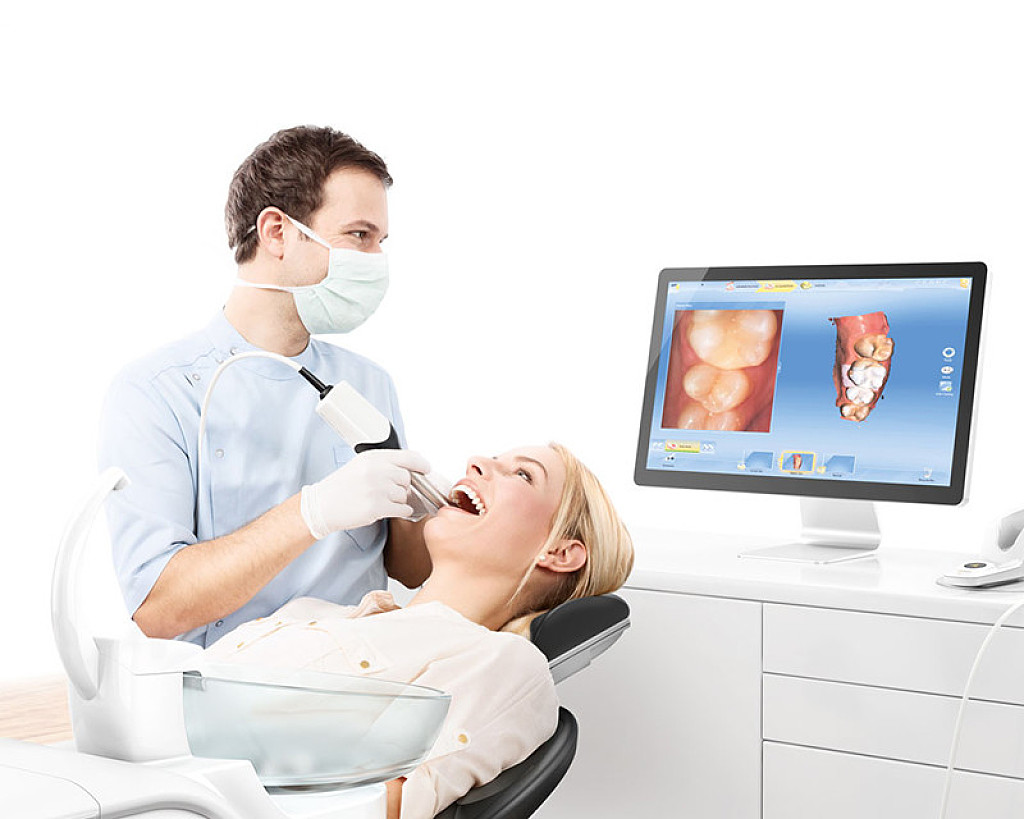
What is CEREC?
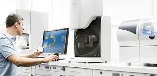
CEREC is the short term for Chairside Economical Restoration of Esthetic Ceramics, or CEramic REConstruction. CEREC uses CAD/CAM (computer aided design/computer aided manufacturing) technology to take impressions quickly and generate a precisely fitted filling so you can leave Discovery Dental sooner.
How can CEREC help you?
One of the biggest advantages of CEREC is its convenience. If you need a crown, inlay, or onlay, you can get your teeth restored during a single trip to Discovery Dental. Traditionally, these procedures require two trips to the dentist.
During the first, the dentist cleans the tooth, makes a mold, and places a temporary restoration onto the tooth. In a couple of weeks, after the permanent restoration is ready, you need to return to the office so that the dentist can remove the temporary fix and place the permanent one.
The CEREC process lets you receive your permanent restoration right here in our Washougal, WA office, so you do not have to live for weeks with a temporary fix and schedule another appointment. In addition, Dr. Dave Stinchfield and Dr. Tom Stinchfield and our team use digital impressions to make a mold for the filling. This is more comfortable and accurate than traditional impressions with plaster.
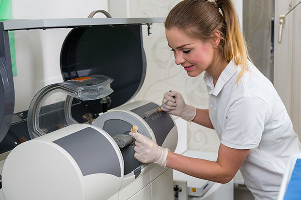
Another benefit of CEREC is that it uses a single block of solid ceramic materials instead of pressed ceramic and metal. CEREC restorations are able to withstand moderate chewing so yours will last for years.
The lifespan of a CEREC restoration is longer than similar work with traditional methods. In addition, the color of CEREC ceramic is closer to the color of your natural teeth, which will make your restoration virtually unnoticeable.
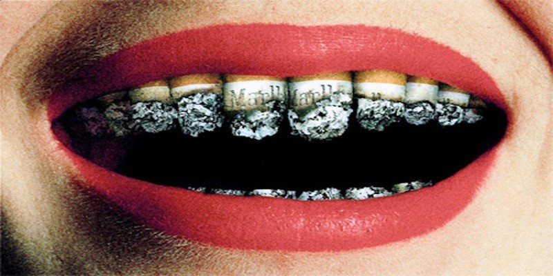
The oral changes from tobacco use range from harmless soft tissue changes to a life-threatening oral cancer.
Your dentist is trained to perform an oral examination to detect tobacco use related abnormalities. Some of the more common of these are discussed below:
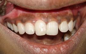
Smoker’s Melanosis
Smoker’s melanosis is increased tissue pigmentation, or darkening, due to irritation from tobacco smoke. Typically this pigmentation occurs on the gingiva (gums) of the upper and lower front teeth. The amount of pigmentation increases with greater tobacco use, and is more common in females; it occurs in 5.0 – 22% of cigarette and pipe smokers. There is no treatment for smoker’s melanosis; however, tissues typically return to normal color in six to 36 months after quitting smoking.
Periodontal Disease
The evidence is overwhelming that smoking contributes to periodontal disease (see Right) and that continued smoking results in a reduced response to periodontal treatment. There is a greater amount of bone loss around teeth in smokers and individuals who smoke are more likely to lose teeth than nonsmokers. It is reported that more than half of advanced gum disease can be linked to tobacco use.
Nicotinic Stomatitis
In nicotinic stomatitis, the hard palate (roof of the mouth) appears white instead of pink, and numerous, small raised areas with red centers are found throughout the palate (see Left). These red areas are irritated minor salivary glands whose duct openings are inflamed in response to the heat from tobacco products. This lesion is most commonly seen in older male tobacco users who smoke pipes but it also can be found in cigar and cigarette smokers.
There is an increased risk for cancer of the tonsils, posterior mouth, and lungs in individuals who develop nicotinic stomatitis from their tobacco use. However, if the individual stops their tobacco use, the appearance of hard palate typically returns to normal within a few weeks.
Gingival Recession and Tooth AbrasionIn addition to the development of changes to the oral tissues, the use of smokeless tobacco can damage both the gum tissue and the teeth in the area where it is held in the mouth. Smokeless tobacco can result in localized gum recession and the exposed teeth often develop dental decay due the sweetener in smokeless tobacco. Unfortunately, stopping the tobacco use does not reverse the gum problem or tooth decay.
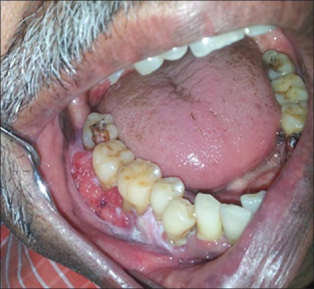
Oral CancerUse of tobacco products is clearly linked to development of oral cancer (see Below). Oral cancers are found primarily in the floor of the mouth (under the tongue), the sides and underside of the tongue, and the soft palate (the back part of the roof of the mouth). The topic of oral cancer in discussed in a separate Patient Information sheet. The most important key to surviving oral cancer is early detection. The importance of your dentist performing a thorough soft tissue examination cannot be overemphasized. The tissue changes in early cancer can be subtle and it is essential for your dentist to perform a through soft tissue examination to detect cancer at an early stage. He or she may want to take a sample of these tissues (biopsy) for diagnosis, or refer you for this procedure. This is the only way to make a diagnosis of oral cancer, and biopsy can also help in determining your long-term outlook.
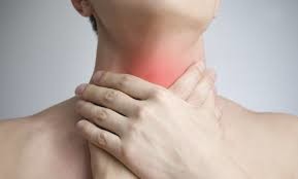
When your neck and/or head is receiving the radiation treatment it needs to fight cancer; your mouth could be indirectly affected by the radiation. And sometimes that could push pause on your cancer treatment — not good. So you do need to be mindful of any possible side effects. You should also integrate your dental team with your cancer care team to keep your mouth healthy before treatment begins and to arm yourself with the knowledge needed if any oral health problems arise.
How Does Head And Neck Radiation Affect Your Mouth?
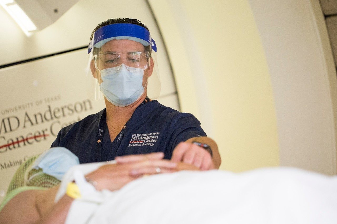
The head and neck radiation should be doing its job and destroying the cancer cells. Sometimes, the radiation can also impact healthy normal cells in your mouth, creating various side effects. There's no rhyme or reason on which side effect touches which patient or for how long. Fortunately, you should be able to see or feel them in your mouth and treat them as necessary. The most common sides effects created from the radiation are:
Difficulty speaking, eating, swallowing
Dry mouth
Cavities
Loss of taste
Sore mouth and gums
Infections throughout the mouth
Jaw stiffness
Jawbone changes
It's crucial that you address any of these that affect you so your cancer treatment can continue as needed.
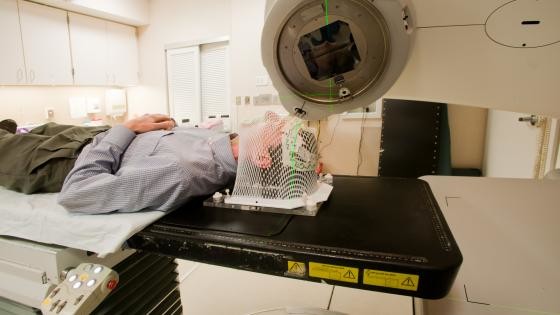
Why And When You Should See A Dentist?
The side effects from your head and neck radiation are a real possibility. But seeing your oral care providers before radiation treatment begins can only help minimize them should they happen. If your oral health is in tip-top shape, the better your chances to stick to your cancer treatment as scheduled. Visit with your dentist at least two weeks before radiation treatment and connect them with your cancer care team, so they're all on the same treatment wavelength. Your dental team will:
Provide a thorough oral exam of your mouth and teeth
Take X-rays
Address any other mouth problems
Show and instruct how to treat and prevent any side effects from radiation
Show and advise how to avoid jaw stiffness by exercising the jaw muscles three times a day with 20 reps of opening and closing your mouth as far as possible
What Can You Do To Keep Your Mouth Healthy?
The first step in keeping your mouth healthy is to — watch your mouth. Look for any noticeable changes visually and try to be aware of pain or discomfort internally. While there is no toothpaste for radiation, you should consider the following tips to keep your overall oral health in good shape.
Prevent dry mouth:
Stay hydrated with water and ice chips
Opt for sugarless gum or candy
Try a saliva substitute to moisten your mouth
Clean your entire mouth:
Brush your teeth (with a fluoride toothpaste) and tongue after every meal and before bedtime with an extra-soft toothbrush (soften bristles under warm water as necessary)
Remove plaque between your teeth using interdental brushes, water flossers, or floss being careful not to irritate or cut your gums.
Mix 1/4 tsp of baking soda and 1/8 tsp of salt with 1 cup of warm, then rinse and follow with a plain water rinse
Check with your doctor or dentist about ill-fitting dentures
Mind what you eat and drink:
Eat foods that are nutritious and easy to swallow with slow bites and sipping liquids while chewing
Choose softer foods like scrambled eggs, mashed potatoes, etc. — and add a sauce, gravy, or liquid for help with swallowing
Try to avoid sharp, hard, crunchy foods, hot, spicy, highly acidic foods, overly sugary foods like candy and soda, all tobacco products, and alcoholic beverages
Contact your team when there's pain:
Take their prescribed pain relief medications as directed