A new paper in Science Advances describes for the first time how minerals come together at the molecular level to form bones and other hard tissues, like teeth and enamel.
The University of Illinois Chicago researchers who published the paper described their experiments -- which captured high-resolution, real-time images of the mineralization process in an artificial saliva model -- and their discovery of distinct pathways that support bone and teeth formation, or biomineralization.
"Until now these pathways, particularly at the early stages when molecules are first starting to organize into a structure, have not been understood clearly," Reza Shahbazian-Yasser, UIC professor of mechanical and industrial engineering at the College of Engineering and corresponding author of the paper.
Shahbazian-Yasser and his colleagues observed that both direct and indirect formations of hydroxyapatite crystals -- the foundation of hard tissues -- can be achieved by local variations in energetic pathways for nucleation and growth.
"The control over the dissolution of amorphous calcium phosphate affects the assembly of hydroxyapatite crystals into larger aggregates," Shahbazian-Yasser said. "Using technology developed at UIC, we found evidence that these pathways coexist simultaneously -- explaining why different groups had reported seemingly different or opposite results. In addition, we now understood how hydroxyapatite materials nucleate and grow on amorphous calcium phosphate templates. The control over the nucleation and growth of hydroxyapatite will aid in developing new drugs and medical treatments to heal lost or broken bone faster or cure tooth cavities."
To capture the images, the researchers used a unique micro-device that made it possible to use electron microscopy with a liquid model. Using this method, the researchers were able to monitor chemical reactions in the model on the smallest scale.
"Our study provides clear, new evidence of how minerals organize and grow into bone materials, and this finding has many important implications for further research on bone or teeth healing," Shahbazian-Yasser said.
"By better understanding these pathways, scientists are one step closer to engineering ways to better treat dental diseases and bone injuries -- like those from traumatic injuries -- or prevent medical conditions that can develop when normal mineralization processes in the body go awry," he said.
Medical conditions caused by dysfunctional mineralization in the body can include everything from a tendency to develop cavities to osteoporosis.
"In the next step, we would like to learn how molecular modifiers can affect the process of biomineralization, which is important to develop effective drugs," Shahbazian-Yasser said.
Pain during or after eating can result from a number of problems, including tooth decay or other dental problems. Make sure you see your dentist every six months for a checkup and discuss any symptoms you’re having.
If you get frequent headaches, especially migraines, you may also notice that certain foods or the act of chewing very crunchy foods can trigger a headache. If this is happening to you, you may need to see a medical professional who specializes in headache treatment.
Many times, however, pain from eating isn’t a tooth or headache issue — it’s a jaw issue. A temporomandibular disorder, or TMD, causes inflammation, swelling, and pain in the jaw and facial area that can get worse when eating, talking, chewing gum or even breathing through the mouth. TMD is often called TMJ, which is actually the name for the affected jaw joint, known as the temporomandibular joint.
Besides jaw pain and pain after eating, people who have TMD may notice:
Jaw popping, clicking, or a grinding, gravelly sound when using the jaw
Regular headaches, especially upon waking in the morning or after using the jawTemple area painPain or stiffness in the back, shoulders, or neck
Inability to freely move or open or close the jaw
Ear pain without an ear infection
Ringing in the ears (tinnitus)
Balance problems or dizziness
TMD is a complex condition — because the TMJ is a complex joint. The bones, muscles, and tendons that allow you to move your jaw in different directions must work together perfectly in order to function without pain or symptoms of TMD. If something throws off this balance, the TMJ may become irritated, swollen, or inflamed. This can quickly lead to TMD symptoms.
Many people think that some soreness or popping of the jaw when eating is normal — it’s not. The jaw should work silently and smoothly when it’s in optimal condition. You should be able to eat without any discomfort or stiffness. If this is not the case, don’t ignore it. Over time, symptoms could get worse. Make an appointment with a medical professional who is educated in TMJ disorders and can guide you through treatment.
Many people are wrongly led to believe that if they have TMD, they should simply “tough it out” until it goes away or that it will never get better. This is not the case!
TMD can be treated in a number of non-invasive and highly effective ways. But, you must see a doctor who is experienced in helping people with this complex condition. At MedCenter TMJ, this is what our doctors do every day.
Every patient at our practice receives a personalized treatment plan designed to get you on the path to healing as quickly as possible. This will likely include a custom orthotic appliance that will help align your jaw in its optimal position. You’ll also receive instructions and plans for stretches, exercises, stress management techniques, and ongoing support. If your pain is severe, we will work with you to find safe, effective pain relief to help you get on with your life. We also help our patients with alternative and natural remedies.
Just like you would not walk on a broken foot until it was healed, you should not excessively use or stress your TMJ if you’re experiencing TMD symptoms. One of the best ways to help rest and heal the TMJ is to follow a TMJ-friendly diet.
A TMJ-friendly diet should include plenty of fruits and vegetables for their anti-inflammatory properties. Proper nutrition and healthy foods can improve TMD symptoms. But, don’t consume fruits and vegetables raw if they are crunchy. Instead, cook them until soft or blend them into a nutritious smoothie.
You’ll need to avoid foods that require extensive chewing, as this will stress an already-irritated jaw joint. Crusty bread, tough cuts of meat like jerky or steaks, chips, and similar foods should be avoided. Try softer options such as very soft, thin breads and meats that have been boiled or slow-cooked for softness. Avoid chewing gum at all times and never chomp on ice, hard candy, or other hard foods!
It's well known that sugary treats, sticky snacks and syrup-laden sodas are bad news for your teeth. But did you know there are some foods that prevent tooth decay? Certain foods either help restore weakened enamel or contain nutrients like protein that can help strengthen it before it wears away. Some foods are better than others when it comes to fighting decay, but for the sake of your teeth, make sure your diet contains some of the following:
1. Crunchy Vegetables and Fruits
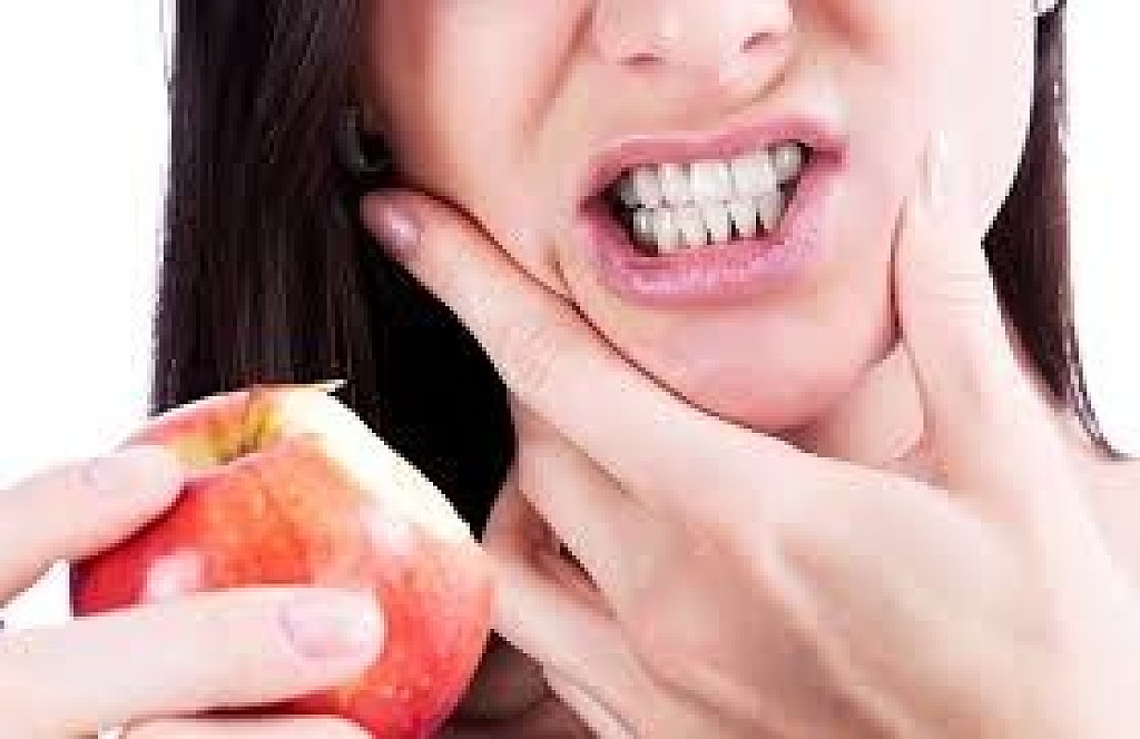
An apple a day might keep the doctor away, but that includes your dentist. Crunchy fruits and veggies are just some of the foods that prevent tooth decay, due to a high fiber content that stimulates the flow of saliva throughout your mouth. Therefore, slices of carrot, celery or apple go perfectly with an otherwise rich main course to help give your teeth a good cleaning.
The saliva you produce when you eat crunchy veggies, in particular, rinses any remaining sugar away from your teeth before it has a chance to attack healthy tooth enamel. Even though some fruits, like apples, are high in sugar, the amount of fiber and water in them is high enough to balance the sugar's effects and do your mouth more good than harm, according to the American Dental Association (ADA).
2. Cheese and Dairy
If you look forward to eating cheese, here's some fantastic news: It happens to be phenomenal for your teeth. Cheese doesn't contain much sugar, but it does carry protein and calcium – both of which are important for the strength and health of your enamel. As a bonus, the Journal of General Dentistry recently found eating cheese actively protects the teeth from cavities. Participants in a study who chewed on cheese for three minutes saw their mouths' pH levels increase over the course of 30 minutes. A low pH indicates higher acidity, and usually increases your risk for tooth decay at less than 5.5. Having a higher pH means a lower risk for decay.
Cheese isn't the only dairy product that is good for your teeth, of course. Research published in the Journal of the American Dental Association showed that drinking milk after eating sweets also reduced the amount of plaque buildup on one's teeth. Like its relative in cheese, milk is a classic source of calcium, which can help remineralize the teeth and minimize decay.
3. Seafood
Fish, lobsters and shellfish can be great for your teeth, and for two reasons. First, they tend to be an excellent source of lean protein, which can help keep your teeth healthy and strong in general. Secondly, most seafood contains fluoride, according to the National Institutes of Health (NIH). This fluoride can help reduce your risk for tooth decay just like the fluoride incorporated in your tap water.
4. Nuts and Other Sources of Protein
If you aren't a fan of seafood or can't eat it due to an allergy, rest assured there are a variety of foods containing the protein that helps your teeth in much the same way. Nuts contain a good amount of protein, as well as calcium and phosphorous, which help strengthen your teeth's enamel. Because nuts are crunchy, eating a handful of them also stimulates saliva production, which can further reduce your risk for tooth decay. Chicken, eggs and other types of lean meat are great for your teeth thanks to their own protein.
What you eat can definitely help to protect your teeth, but a healthy diet isn't a replacement for a great oral care routine at home. Along with eating tooth-healthy foods, remember to brush twice a day using a fluoride toothpaste and floss daily. See your dentist twice a year for cleanings and a checkup so any problems get treated before they become a bigger deal than they need to be.
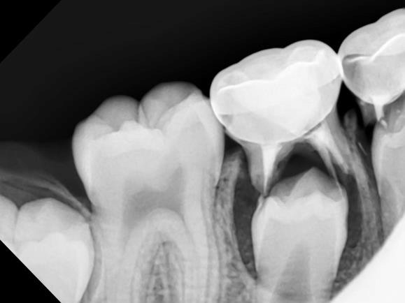
A tooth filling is where material (such as porcelain inlay/onlay or composite) is inserted into a damaged or decayed tooth to restore its original shape and function. This can be either a dental inlay (fills centre of tooth) or onlay (more extensive than an inlay and can cover up to the entire biting surface of a tooth).
Fillings are most commonly used to prevent further damage to a tooth when decay is present. By removing the bacteria and closing the affected area off, no further tooth decay can occur.
The process of getting a tooth filling
Once your dentist has assessed your case and determined a tooth filling is the best solution, you will be booked in for your next treatment. In general, a filling treatment can last anywhere roughly between 30 minutes to an hour to complete all the necessary dental work.
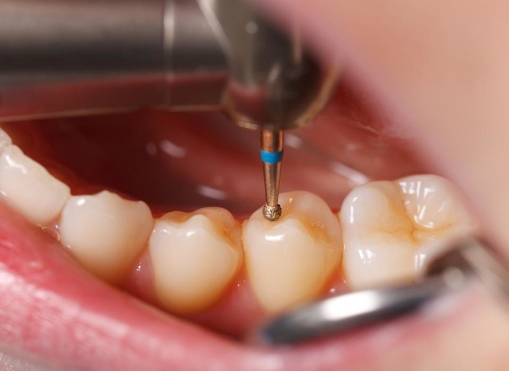
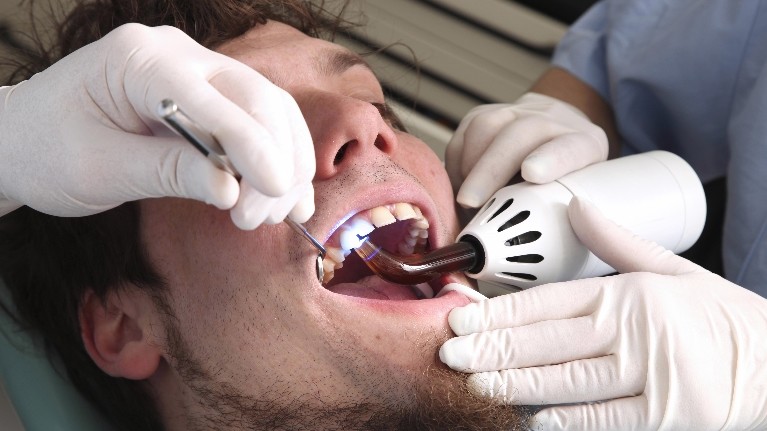
Most dentists will prepare the area with numbing spray/gel before local anaesthetic is used. This creates a more comfortable experience for patients by lessening the feeling of the local anaesthetic needle.
Once the area is completely numb, the dentist will remove any decay with a drill and seal the area with a filling. To maintain proper bite function, the dentist will check with the patient to ensure that their bite feels normal. If necessary, part of the filling will be filed down to restore the natural bite.
After the treatment, your mouth will be numb for a few hours. During this time, it is not recommended to chew on the numb side of your mouth.
Fillings are a relatively straight forward procedure. However, it is good practice to keep your dentists number on hand in case you experience any discomfort.
Types of fillings
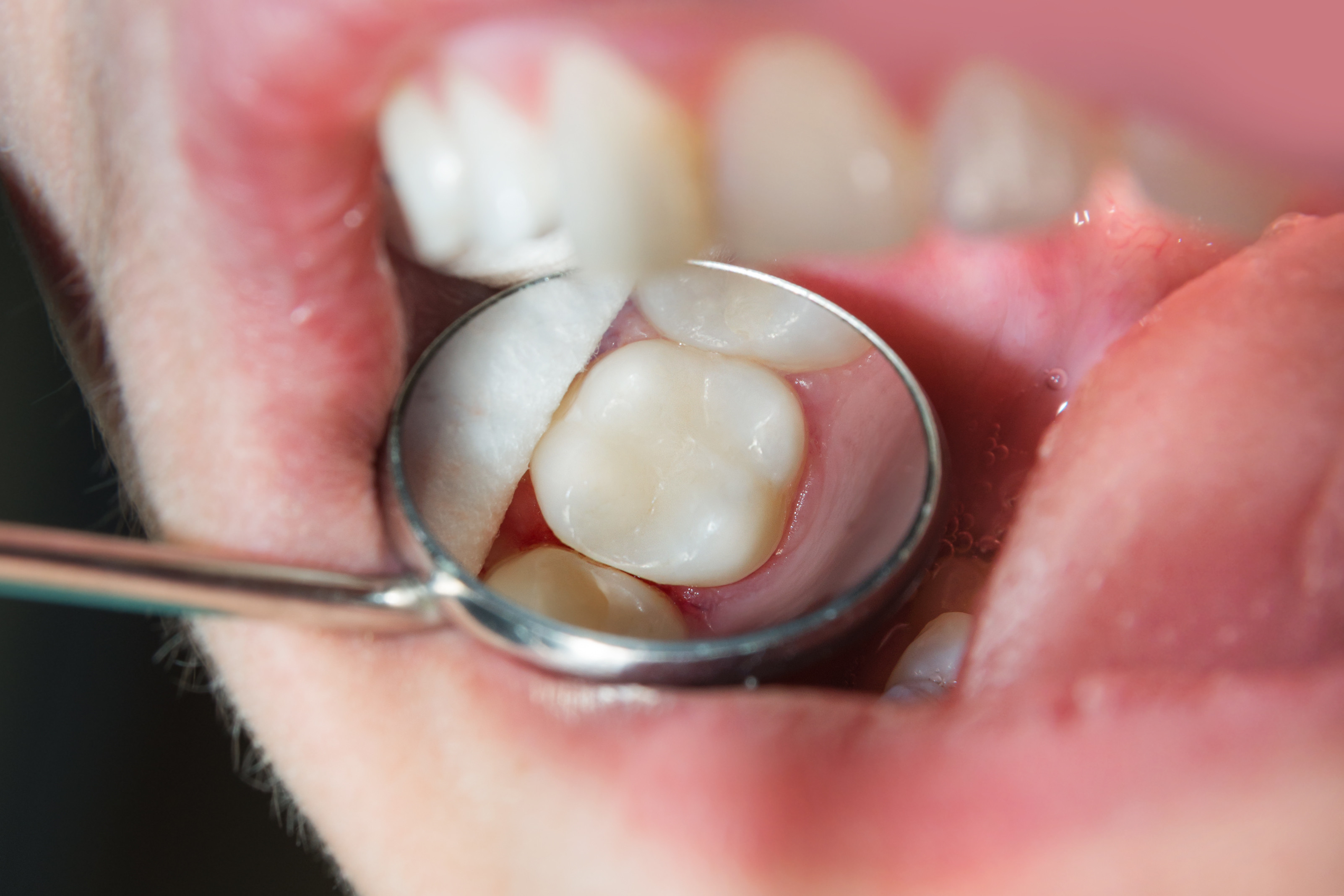
Porcelain fillings – inlay/onlay are tooth coloured and are created to match the current aesthetics of teeth. A porcelain filling is virtually indistinguishable from other healthy teeth and can last up to 20 years.
Composite fillings are similar to porcelain fillings; however, they can last 5-10 years when applied to teeth.
Gold fillings need to be created in a lab to fit the tooth cavity and are then cemented into place. They are the most expensive type of filling and require multiple visits. Gold fillings are well received by the surrounding gum tissue and can last for more than 20 years.
Amalgam fillings were the most common, cheapest filling available. However, amalgam fillings contain mercury and are not commonly used in Australia. Moreover, this type of filling can expand and contract in heat which can cause teeth to crack. For this reason, we do not offer amalgam fillings at Coastal Dental Care. Instead, we offer to replace old amalgam fillings with healthier tooth coloured fillings.
Aftercare for tooth fillings
It is safe to eat normally almost instantly after receiving a new tooth coloured filling. Although, it is important if you have had local anaesthetic not to chew on the numb side. When your mouth is numb or partially numb, you have a greater risk of biting your tongue or lip.
Also, be careful of hot drinks as the anaesthetic reduces your ability to gauge the heat of the liquid and you might burn yourself.
If you have had a deep filling, you might continue to experience pain from the inflamed area. This should fade away within a few days as the inflammation reduces. If pain continues, please consult one of our Gold Coast dentists.
Keep in mind, a healthy diet, regular brushing and flossing and regular 6-monthly check-ups and clean can help to reduce your risk of developing tooth decay in the future.
Many common dental health concerns are affected by genetics, which is why your dentist will ask about your family history at appointments. While a genetic predisposition doesn’t mean you’ll definitely get the condition, it does affect your risk for it, and your dental care should be adjusted to account for it. Below are a few common dental issues that you should watch for.
A Guide to Dental Conditions That Are Genetic
1. Gum Disease:
Gum disease, or periodontal disease, has been linked to a family history of the condition. If one or more members of your family have had gum disease, you’re more likely to get it, even if you brush and floss daily. A genetic test can be done to analyze your risk, along with taking your family history, so that defensive measures can be taken, like using plaque-fighting products and having more frequent checkups from a dentist.
2. Cavities:
Tooth decay is caused by bacterial excretions that erode your enamel, and it can lead to tooth loss and treatments like dental crowns. It’s generally associated with poor diet and oral hygiene, but scientists have shown that it could also occur due to genetic susceptibility to the bacteria in your mouth. Family history and genetic testing can determine your risk for tooth decay, and preventive measures like sealants, fluoride treatments, and rigorous oral health can minimize the risk.
3. Jaw-Related Issues
The size of your jaw is determined by genetics, and if your jaw size is off, it can lead to dental issues. For example, if it’s too small, then crowding and crooked teeth are common. If it’s large, gaps and misalignments will occur. Small jaws also have a higher risk of impacted wisdom teeth, as they don’t have space to emerge properly.
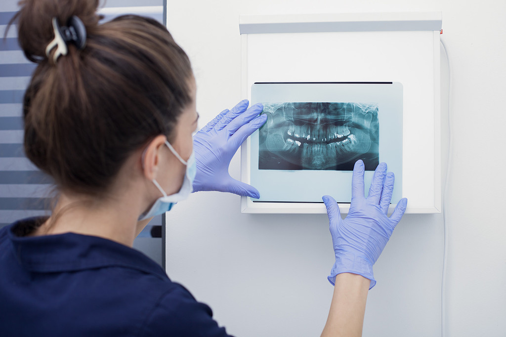
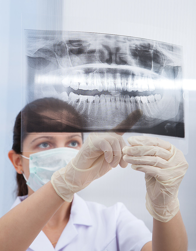
If your family history includes the need for orthodontic solutions or impacted wisdom tooth removal services, chances are that you will need them, too. X-rays or digital imaging can determine the positioning of wisdom teeth and the likelihood of issues coming up, while braces will help correct misaligned teeth.
Clinical hypnosis seems to bring advantages to dentistry for it helps neutralize indefensible nervousness and phobia in patients. Dentists who make use of hypnosis are more able to relieve their anxious patients’ pain and fear. This article provides an insight into the advantages of hypnosis as a therapy, and explores its applications in dentistry.
Dentists who use hypnosis regularly in their clinical practices appreciate a variety of significant advantages. There are many and varied applications of clinical hypnosis in dental clinical practice. Dental applications of hypnosis include relaxation, relief from fears and anxieties, reduction in both the perception and severity of pain during procedures, control of bleeding and salivation (both for increasing and decreasing flow, as needed), control of bruxism (tooth grinding), finger-sucking, and other habits, and promotion of behavioral modifications associated with optimization of oral health.
Amongst a lot of options for behaviour management and modifications, hypnotics is one of the oldest and non-invasive way to control dental anxiety in children, adult as well as in geriatric patients and hence get a better treatment result and a good compliance and satisfaction of the patient.
Use of hypnosis in the dental practice
Hypnosis can bring considerable relief to anxious patients and make it easier for the dentist to do their job but it is particularly implemented in order to help patients relax. As relaxation raises the pain threshold, requirement for local anaesthesia is reduced. And even if it is necessary, it is better tolerated. Therefore, the use of hypnosis as a general relaxation strategy is certainly possible and there are reports in the literature of its use in both adults and children.
Moreover, clinical benefits can be derived from hypnosis such as the control of dentophobia, abnormally active gag reflex, trigeminal neuralgia pain, benign chronic orofacial pain, temporomandibular joint dysfunction (TMD), adaptation to dentures, behaviour modification, like thumb sucking, bruxism. Additionally, hypnosis can control salivary flow and bleeding. Xerostomia and haemostasis can be produced through hypnotic suggestions, such as visualizing being in a desert on a hot day and noticing how dry the mouth becomes because of lack of water. Mental imagery of the ligation of a bleeding vessel can be used to decrease bleeding after soft tissue surgery.
Hypnosis in children and adolescents is possible, but, much harder to administer than in the adults. It is also true that not everybody is susceptible to hypnosis, as it is apparent that this phenomenon has also some association with genetics and brain structure.
Current methods of hypnosis
A distinction arises between a deep type of hypnosis and a light one and both have different applications. Deep hypnosis takes long so it is not apt for regular dental practice; however, it is required for analgesia and behaviour modification. On the other hand, the ‘light’ state is easier and faster to attain and is used in hypnodontia on a daily basis; for instance, to relax a nervous patient in a matter of minutes.
Dentists have to use positive suggestions managing patients. Words or actions that inspire trust in the dentist will relieve the patient’s anxiety and fear. Informal hypnotic methods include the use of utterances like “you will feel quite comfortable” or “you will like the results” and this can be a powerful technique of patient management. Suggestions can be categorized into two broad types: direct and indirect.
Direct suggestion involves straightforward statements that are clearly understood by the hypnotic subject. An example would be: “don’t move your head because you won’t be able to before finishing.” Indirect suggestion uses indirectness in addressing the subject in a form of covert hypnotic statements. That is to say the subject is hypnotized without their knowledge.
Conclusion
Hypnosis has many uses within the dental field, ranging from simple relaxation of the anxious patient to complete analgesia for surgery. Clinical hypnosis can be an incredibly valuable tool in dentistry.
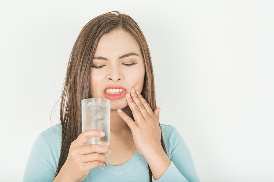
If you feel sudden tingling sensation in the mouth upon taking something hot or cold, you might have tooth sensitivity.
Tooth sensitivity, also known as dentinal hypersensitivity, is a common dental issue in the age group of 20-50 year old. This is a condition wherein the teeth may become extremely sensitive to hot or cold temperatures. Therefore, you would feel a sharp sensation in the teeth while consuming hot or cold food and drinks.
What Causes Tooth Sensitivity?
The problem is quite common these days and following are some of the reasons for the same:
Recession of gums owing to gum disease or rough brushing practices
Wearing off of enamel as we age, due to grinding of teeth, or increasingly frequent brushing
Exposure of the soft inner part of the tooth called dentine, which lies between the enamel and gums
How to Know If I Have Tooth Sensitivity?
As mentioned earlier, your dentist would be the best judge of your situation.
Here are a few common symptoms as reported by people who have experienced tooth sensitivity:
A sharp sensation while consuming cold food and beverages
While consuming hot food and beverages
Consuming sweet or sour food items
Breathing cold air through the mouth
Slight pain while brushing teeth
Tips for handling tooth sensitivity:
Dr. Sahakyan suggests a few simple remedies to help deal with tooth sensitivity. You may try these but most important, if the problem still persists, visit your Glendale dentist soon.
Avoid Aggressive Brushing – Sometimes brushing too hard or too frequently may lead to sensitivity. Try changing your toothbrush at the earliest. Using a soft-bristle toothbrush just about twice a day can help. Don’t brush too fast or roughly on your teeth.
Change Your Toothpaste – Switching to a toothpaste specially designed for sensitive teeth can also help solve the problem.
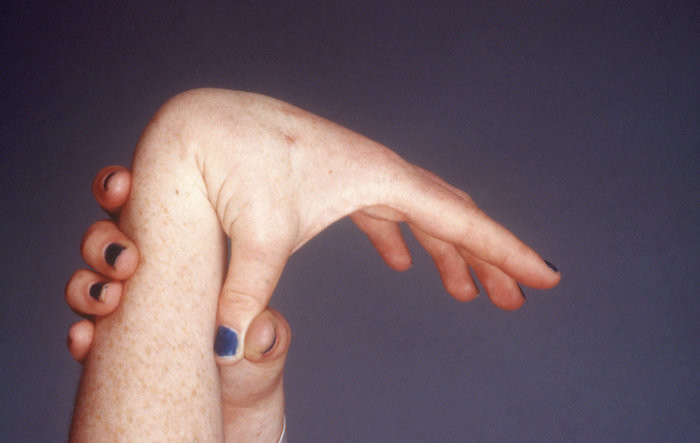
Ehlers-Danlos syndromes (EDS) are a group of rare inherited conditions that affect connective tissue.
Connective tissues provide support in skin, tendons, ligaments, blood vessels, internal organs and bones.
“When contemplating orthodontic treatment on a patient with Ehlers-Danlos syndrome (EDS), there are a number of special precautions to be taken. Extreme joint hypermobility in many EDS patients often leads to chronic dislocation of the temporomandibular (jaw) joint.
This makes the placement of complex orthodontic appliances very troublesome for the patient and the clinician. In addition, the oral surgeon must be extra cautious to prevent a dislocation of the mandibular condyles (lower jaw joint) when performing a surgical procedure in preparation for orthodontics. Because of tissue repair problems in EDS, there may be slow healing after dental extractions, followed by soft tissue scarring. The orthodontic appliance used on an EDS patient should be very smooth and relatively simple in design. The oral mucosa, or mouth lining tissues, are very fragile, liable to injury and particularly vulnerable to sharp objects such as orthodontic appliances (braces) or partial dentures.
The dental anatomy of the posterior teeth occasionally have high cusps and deep fissures. The roots may be dilacerated, (stunted, bent, fused or twisted in shape). The pulps may become partly obliterated by the pulp stones in the crown portions of the pulp, making root canal treatment difficult. The dentin may have an unusual pattern and abnormal fine structure because of an aberrant collagenous dental crown anatomy. Thus, there may be a right to left or upper dental arch to lower dental arch tooth size discrepancy (difference) making ideal dental interdigitation very difficult.
Tooth movement might be expected to be more rapid for a constant appliance activation because of the collagen cross linkage defect. The mobility of teeth during tooth movement may be greater than normal. This may be caused by stretching, tearing and slow repair of the fibers. Similarly, the gingiva (gums) may be more prone to inflammation and possible recession. There have been reports of early onset of some periodontal defects (gum and tooth support). The old EDS type VIII, which is similar to the Classical type, in particular, is characterized by extreme periodontitis which can be quite debilitating.
With the added dental mobility of the teeth, slowed repair processes and poor organization of tooth supporting tissue collagen, the need to wear retainers long after completion of the case may be greater. Although anatomic defects in the root morphology have been described in EDS, the detailed molecular composition of the dentin has not been studied. If changes do exist, root resorption as a side effect of orthodontics could be a problem. This has not been demonstrated clinically, however.
EDS is a connective tissue disorder which may have many effects on the dentition of the patient. With suitable understanding of the underlying disease manifestations and appropriate precautions by the orthodontist, orthodontic treatment can be accomplished with the minimal undesirable side effects.Dental Manifestations of EDS– Hypermobile temporomandibular joint (TMJ); high incidence of subluxation.– Fragile oral mucosa.– Early onset of periodontal defects.– High cusps and deep fissures on the crowns of teeth.– High incidence of enamel and dental fractures.– Stunted roots or dilacerations.– Coronal pulp stones.– Aberrant dentinal tubules.– Pulpal vascular lesions and denticles.– Teeth move readily in response to orthodontic forces.– Orthodontic retention easier to accomplish.
(12/27/2020) by We Strength Flexibility Health EDS
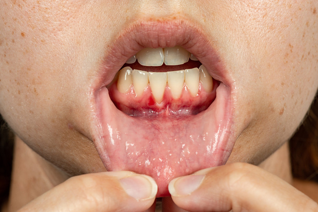
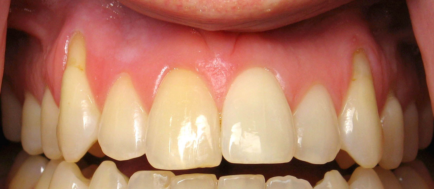
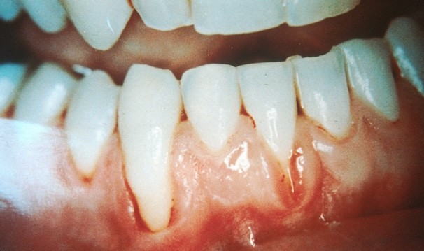
Gum recession is when the margin of the gum tissue surrounding the teeth wears away, or pulls back, exposing more of the tooth, or the tooth's root. When gums recede, gaps can form between the gum and tooth, allowing disease-causing bacteria to build up. If left untreated, the surrounding tissue and bone structures of the teeth can be damaged.
Receding gums is a widespread dental condition. Most people aren’t aware that they have receding gums since it occurs gradually.
Receding Gums Symptoms:
As receding gums progress over time, you may notice the following symptoms:
Long Teeth.- One symptom is the visible lengthening of the teeth. When gums recede because of periodontal disease, the teeth have the appearance of being much longer than normal.
Exposed Roots.- Exposed roots are another symptom, and can be extremely sensitive and uncomfortable. They are often a sign of periodontal disease or can be attributed to brushing overly aggressively with a toothbrush with hard bristles.
Loose teeth.- When suffering from receding gums, you may notice loose teeth, attributed to the bacteria and periodontal disease under the gums around the teeth. As receding gums worsen, the gum pockets deepen due to loss of attachment structure.
Causes of Receding Gums:
Numerous factors can cause your gums to recede, including:
Periodontal diseases.- These are gum infections, caused by bacteria, that destroy gum tissue and the bone that holds your teeth in place. Periodontal disease is the main cause of gum recession. The early stage of periodontal disease is not often painful, therefore symptoms often go unnoticed. Left untreated though, early symptoms can develop into periodontitis.
Early stages of gum disease can be seen with minor symptoms that include:
Red, swollen, or purple gums
Gums that feel tender to the touch
Bleeding gums
Chronic bad breath
Genetics.- Some people are more susceptible to periodontal disease. Studies show that 30% of the population may be predisposed to gum disease, even if they take good care of their teeth.
Brushing too hard.-If you brush your teeth too aggressively or incorrectly, it can cause your tooth’s enamel to wear away and your gums to recede.
Poor dental care.-Inadequate brushing, flossing, and rinsing with antibacterial mouthwash, can make it easy for plaque to turn into tartar, a hard substance that forms on and between your teeth and can only be removed by a professional tooth cleaning.
Hormone levels.-Changes in estrogen levels over a woman's life, like puberty, pregnancy, and menopause, can make gums increasingly sensitive and vulnerable to gum recession.
Tobacco products.-Smokers, and other tobacco users, are more likely to develop sticky plaque which can cause gum recession.
Grinding and clenching your teeth.- Clenching or grinding your teeth can exert too much force on the teeth, causing gums to recede.
Crooked teeth or a misaligned bite.-When teeth don’t come together evenly, too much force can be exerted on the gums and surrounding bone, allowing gums to recede.
Receding Gums Treatment:
Mild gum recession can be treated by a professional deep cleaning in the affected area. During the deep cleaning, plaque and tartar is removed and the exposed root area is smoothed over, making it more difficult for bacteria to attach itself. Antibiotics can also be used to kill any remaining bacteria.
If a deep cleaning is not sufficient to treat the condition, because of excess loss of bone and deep pockets, receding gums surgery may be required.
Recently, there has been some talk about the future of dental filling. These composite restorations could be moving to include a new type of material. Our current dental fillings work well but there is always room to improve. Eventually, we could even have special fillings made of glass!
That’s right, glass dental fillings. Sounds crazy, we know but new research shows that glass could be the solution to an even better and more effective dental filling. When it comes to glass dental fillings, we aren’t talking about just any old glass, this special glass is “bioactive” and has the ability to prevent further tooth decay. This could be a total game changer. Dental fillings doubling as a tooth decay prevention.
Most fillings today are created with composite or metal. These are both excellent choices but even these also come with a few drawbacks. Composites are great because they are natural-looking and are made to resemble a person’s natural tooth color. With composite fillings, people will not even notice you have a filling. This great for people who want to be confident in their smile. The downside to composite fillings is that they are not as strong as a person’s natural tooth and the material becomes weaker overtime. A composite filling has a lifespan of about five to seven years.
Metal fillings are another great option for a dental filling. Metal usually has a long lifespan making them the desired choice for tooth that can’t be seen. The lifespan for this filling ranges from ten to fifteen years. The downfall to metal fillings is that overtime it might begin to corrode.
Everyone’s teeth are different and every dental filling is diverse. Things like dental hygiene and diet will always play a role in the lifespan of dental fillings.
The number one reason that fillings need to be replaced is time. The dental material wears down and replacements are needed. In addition, many people experience secondary tooth decay where the tooth continues to decay where the tooth meets the filling. Other things like, teeth grinding and tooth injuries can cause the tooth filling to need replacing.
Believe it or not, glass fillings could be the next trend of the dental world to help fixes these issues. The special bioactive glass has a huge advantage by increasing the lifespan of fillings and even preventing further decay. Research has proven that the unique glass helps slow secondary tooth decay by decreasing bacteria. This special glass is made with silicon oxide, calcium oxide, and phosphorus oxide giving the look of powdered glass. Once the filling is in place, the body will react unlike current fillings. This will help prolong the lifespan of the filling. Researchers have even discovered that cavity-causing bacteria avoids the bioglass. This special bioglass has been used for healing bones for years and could be making its way into the world of dental fillings. Hang tight and you could see this in South Charlotte Dentistry.
Researchers at Oregon State University are leading the discoveries on the bioglass phenomenon. There is no question about what people want most when it comes to dental fillings, something that is long lasting. These engineers were determined to find just that, which led to the bioglass. Their research has been published in Dental Materials, a dental journal published by the National Institute of Health. Composites that contain up to 15 percent of the bioglass are currently being researched and the hope is to eventually incorporate more bioglass into dental filling material.
Bioglass isn’t the only new dental filling being researched. Other studies have found that graphene oxide may also be effective as a dental filling material. This material has the outstanding benefit of not corroding. It is stronger than steel and is very durable. The author of the study, Dr. Stela Pruneanu, has stated that graphene is 200 times stronger than steel. Wow! That would definitely reduce the need to get a filling replaced. Dr. Pruneanu goes on to explain the goal of the study, which is to incorporate graphene into dental materials. They want to do this to reduce corrosion and improve the properties of current dental materials. The study has already seen good results and they are moving onto a different stage in the study. These scientists will begin studying how compatible graphene oxide fillings are with teeth and how the new material will interact with human cells. The material is still being tested in dental materials but incorporating this material could be a huge step forward in filling materials.
These new dental fillings, have not been approved for usage yet, but this could be a noteworthy invention for the future of dentistry.