Chlorine is a necessary and effective way to kill harmful bacteria in drinking water and swimming pools. But you may be wondering, "is chlorine bad for your teeth?" We'll break down the potential risks of chlorine on your oral health and let you know how to protect yourself so you can continue smiling.
Why is Chlorine Used in Water?
According to the American Chemistry Council, before chlorine was used in drinking water to kill disease-causing germs, waterborne diseases killed thousands of people every year.
In pools and hot tubs, chlorine and pH are the first defense against hazardous germs that can make you sick with recreational water illnesses that cause symptoms like: Diarrhea, Skin rashes, Ear pain, Coughing, Congestion And eye pain.
The chlorine included in your tap water is generally not enough to cause any dental problems, but soaking in a backyard jacuzzi or doing laps at your local swimming pool regularly could have negative effects on your tooth enamel.
What Are the Effects of Chlorine on Your Teeth?
Chlorinated pools and hot tubs contain pH levels that can cause enamel erosion on your teeth. Of course, it's unlikely that you swim with your mouth open (if you do, don't), but water tends to seep into your mouth occasionally. A few visits to the local pool a year is unlikely to have any adverse effects. Still, if you swim laps daily or soak in a hot tub every night, the possibilities of enamel erosion on your teeth are real – particularly if you over-chlorinate your pool. The CDC recommends the pH levels of treated water to be between 7.2 and 7.8. The free chlorine concentration should be at least 1 part per million in pools and at least 3 parts per million in hot tubs.
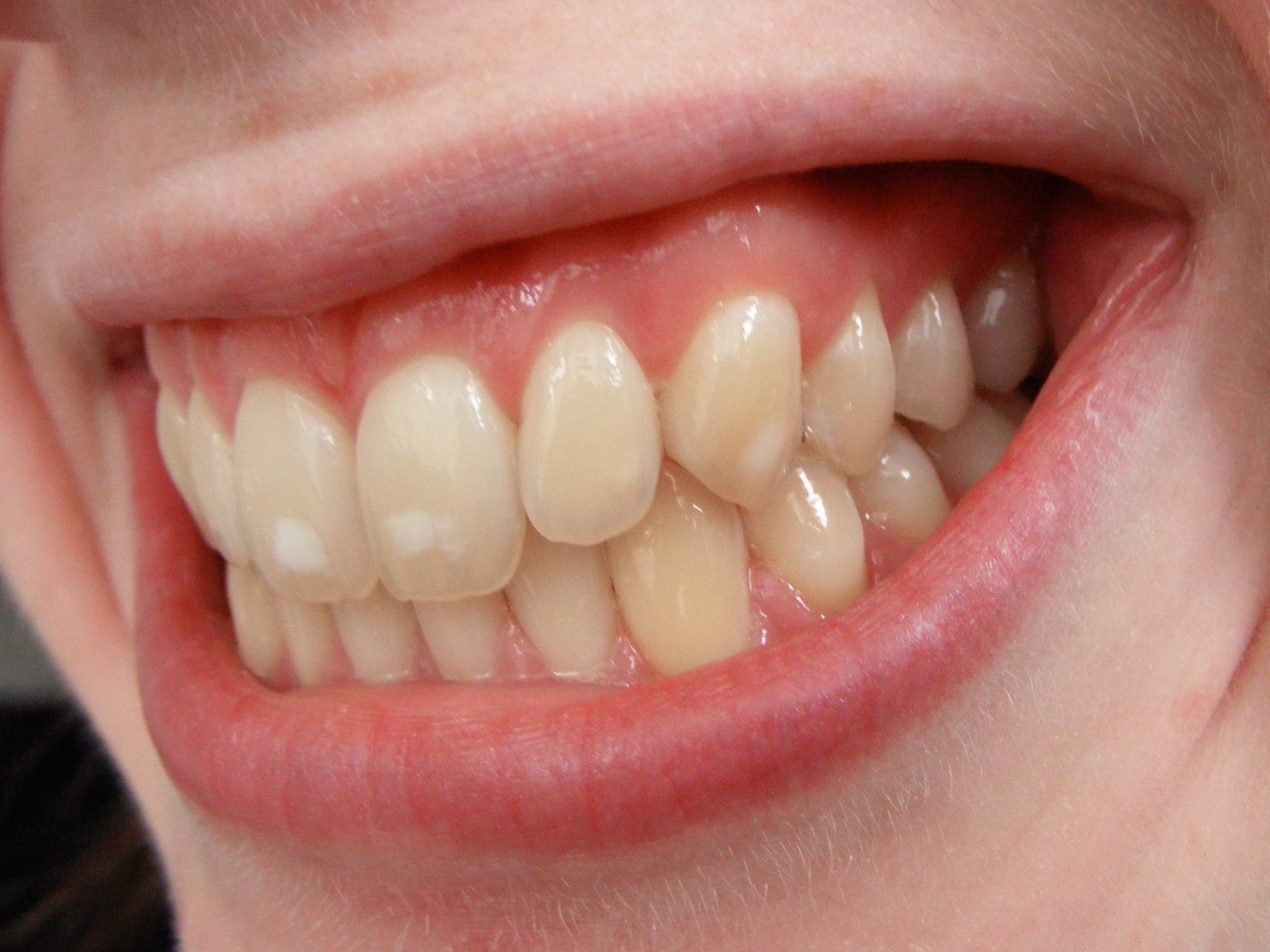
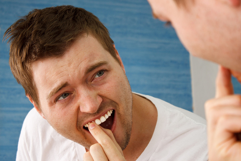
If you notice any of the following symptoms after frequenting chlorinated bodies of water, your tooth enamel may be wearing away (what's called swimmer's calculus). Your teeth may:
Become discolored.
The edges of your front teeth may look transparent.
In later stages, you may feel extreme dental sensitivity when consuming hot or cold foods.
Learn more about how enamel erosion can affect your teeth.
How Do You Protect Your Teeth from Chlorine?
The pH level of water is invisible to the naked eye, so here are some tips to help you know if it's safe to take a dip:
When in a public pool or on a tropical vacation, take notice of pool linings, railings, and ladders. Pool water that's too acidic will eat away at these surfaces. If you notice spots of erosion, the water may do the same to your teeth, so consider skipping your swim or consider swimming elsewhere (perhaps a natural body of water).
Pool pH strips are common in local recreational supply stores and allow you to test the water before wading in.
If you're a homeowner, you might attempt to save money by maintaining your own backyard pool – but this can be tricky. Check your pool's pH balance once a week at a minimum, and budget permitting, hire a specialist to examine it upon your first use.
By taking these precautions when you swim in chlorinated pools and limiting how often you swim or relax in chlorinated water, you can significantly reduce your risk for enamel erosion. If you're an avid swimmer, be conscious of how much water gets in your mouth. And practice good oral hygiene for an even greater chance to withstand the effects of chlorine. Brush at least twice a day, and don't forget to brush your tongue. Consider using a toothpaste that helps replenish natural calcium to strengthen your tooth enamel. And be sure to see your dental professional for regular appointments so they can catch any developing erosion early. When you do all of this, caring for your oral health should go swimmingly.
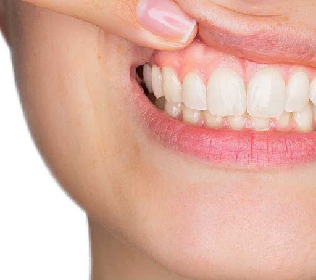
In some cases, changes in your mouth can be an early sign that something is going on elsewhere in your body. Take your gums, for instance. Healthy gums are firm and pink to brown in color depending on your ethnic background. If the color of your gums change or if you develop pale gums, it can be a sign that something's up.
Several conditions can affect the color of your gums. If you are concerned about the look of your gums, your dentist or doctor can help to diagnose the issue and work with you to improve your overall health. Here are a few possible causes of pale gums.
1. Anemia
An article in Periodontology 2000 notes that people with anemia may have pale tissues in the mouth. When a person has anemia, their blood can't deliver an adequate amount of oxygen to the rest of their body, as the National Institutes of Health (NIH) notes. Often, people who have anemia don't have enough iron, which your body needs to make hemoglobin — the protein that gives blood its bright red color.
Several things can trigger anemia. Some people develop it after losing a lot of blood, explains the NIH. Others may develop anemia because their body destroys red blood cells at a higher rate or has trouble producing red blood cells.
Having pale gums isn't the only sign of anemia. Other symptoms include fatigue, unusual heartbeat, weakness and pale skin, according to the Mayo Clinic. Your doctor can run blood tests to look at your red blood cell count and hemoglobin levels before making a diagnosis. They may also perform a test that examines the shape and size of your red blood cells, as the Mayo Clinic points out.
Treatment options for anemia depend on the type. For instance, if you have anemia because your iron levels are low, your doctor might prescribe an iron supplement.
2. Kidney Disease
The kidneys have two jobs: They filter your blood, and they produce urine. When something's wrong with your kidneys, they can't filter waste well. As the National Institute of Diabetes and Digestive and Kidney Diseases (NIDDK) notes, people with certain conditions, such as high blood pressure or diabetes, have a higher risk of developing kidney disease.
As for its effect on gum color, a study published in the Journal of Clinical and Experimental Dentistry compared the gums of 30 patients with kidney disease to a control group of 30 people without kidney disease. No one in the control group had pale gums, while 42.2% of those who had kidney disease did.
Other symptoms to look for if you are concerned about kidney disease are swelling, changes in urination, itchy skin and weight loss, notes the NIDDK. If you have concerning symptoms, schedule an appointment with your doctor, especially if you have diabetes or high blood pressure.
According to the NIDDK, your doctor is likely to order a urine test and a blood test. The NIDDK also explains that treatment of kidney disease often involves taking medication to slow the progression of the disease, monitoring the condition of your kidneys and properly managing other conditions, such as diabetes.
3. Menopause
In some cases, changes in your gum color can also be connected to menopause. The American Academy of Periodontology notes that menopausal gingivostomatitis affects a small percentage of women. One of the signs of menopausal gingivostomatitis is a change in gum color, such as the gums turning pale.
Weight gain, changes in your sleep, mood swings and hot flashes are among the other signs of menopause, according to the National Institute on Aging. Your doctor can provide support and advice to help you cope with body changes associated with menopause.
The appearance of your gums can be a key indicator that something may be going on in your body. If you're concerned about pale gums or other changes in your mouth, don't be shy about discussing your concerns with your dentist or doctor. They can help you figure out what's going on and recommend the proper treatment for your situation.
Sensitive teeth, sometimes called dentin hypersensitivity, is often the result of overzealous tooth brushing, a stiff-bristled brush or use of an overly-abrasive toothpaste, which can wear away tooth enamel over time to reveal the tooth's inner layer of dentin. Tooth enamel can also erode due to acid reflux, bulimia or a similar condition that causes stomach acid to enter the mouth. It may come from excessive amounts of acidic foods and beverages, as well.
Why Just One Spot?
Keep in mind that grinding or clenching your teeth can cause nerve irritation – in localized areas if you have an imperfect bite – as can recent editions of tooth whitening treatment. And if periodontal disease has caused your gums to recede due to an abscess in one particular area (more on that below), the exposed root can be just as sensitive to hot and cold.
Treatment Options for Dentin Hypersensitivity
Having ruled out other causes for your sensitivity, your dentist may recommend desensitizing toothpaste such as Colgate® Sensitive Complete Protection, which helps to seal off the dentin tubules (connected nerves) that cause the discomfort. It usually takes at least a month of regular use before you notice results.
In the meantime, be willing to accept an in-office desensitizing treatment or prescription fluoride gel, which you can apply to the sensitive areas after brushing. And of course, your dentist will work with you to correct any bad habits that may have contributed to the problem.
Tooth Decay
If one tooth in particular bothers you, your dentist will examine the tooth in question and ask you to describe your symptoms. Most likely, he or she will take an X-ray to determine if a few common conditions could be the problem – one of which is tooth decay.
In this case, acids produced by the bacteria built up against the tooth can eventually dissolve its enamel, exposing the dentin layer. And because dentin is filled with tiny nerve endings, you may experience temperature sensitivity and pain when biting down. Once your dentist removes the decay and fills the tooth with either an amalgam or tooth-colored filling, you should be pain free. A full-coverage crown might be needed for more stability and longevity.
Loose or Broken Filling
Unfortunately fillings don't last forever, and when they break or become loose, you may experience sensitivity due to bacteria getting underneath, causing the tooth to decay further. Luckily, this problem is easily remedied with a new filling or crown.
Cracked Tooth
A cracked tooth may not be visible to the naked eye. It may not even show on X-rays, making a diagnosis difficult. However, the American Association of Endodontists (AAE) says a common sign of a cracked tooth is a sharp pain when biting down, but one that disappears after releasing that bite. Cracks involving a break around a filling can be repaired with a new filling or crown, but when a crack extends into the pulp of the tooth, you may need root canal treatment before a crown can be placed. Ultimately, a crack that extends below the gumline and into the root of the tooth will need to be removed.
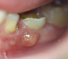
Abscess
An abscessed tooth occurs when the pulp of your tooth – which is made up of nerve and blood vessels – becomes infected. The American Dental Association (ADA) explains that symptoms can include fever, persistent pain and facial swelling. Usually, there's pus-filled swelling at the root tip, which drains periodically and gives you a bad taste in your mouth. You will need root canal treatment to save any tooth that has abscessed.
Recent Treatment
Dental procedures, like removing deep decay or preparing a tooth prepared for a crown, can inflame the nerves within the pulp tissue. This can cause a temporary sensitivity to hot and cold, but it usually dissipates after a week or two.
Whether you have one sensitive tooth or several, it's wise to see your dentist right away. Early diagnosis and treatment can ensure that small problems won't progress into more serious dental complications or the loss of a tooth.
Your tooth enamel is the toughest substance in your whole body, but it's not invincible. Tooth enamel can become worn away by dietary acids (like sodas or juices) or stomach acid, resulting in dental erosion. The American Dental Association explains that acid erosion can make your teeth more susceptible to decay; however, it can also lead to changes in the appearance of your smile that may make you feel self-conscious.
Here are three cosmetic consequences of eroded teeth. Don't worry too much though; it's treatable and preventable!
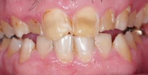
1. Discolored Teeth
Discolored, yellow teeth are a clue that your enamel is being eroded. Your enamel is white, but the tissue underneath the enamel – the dentin – is pale brown. As the enamel thins due to acid erosion, more of the dentin's color will show through, which gives your teeth a yellow appearance.
2. Translucent Teeth
Normally, teeth should be opaque, but if you're experiencing enamel erosion, you may notice that the edges of your front teeth are becoming translucent. Enamel is a semi-translucent substance, but if it becomes worn and thin due to acid, more light is able to pass through it.
3. Rounded Teeth
A change in the shape of your teeth is another possible sign of enamel erosion. You may notice that your teeth look shorter or more worn down, and you may notice that the edges of your teeth are rounded. These shape changes are caused by the loss of your enamel.
Treatments for Dental Erosion
While the cosmetic effects of dental erosion can be unpleasant, your dentist can help you restore the look of your smile. If the erosion is minor, your dentist may recommend strengthening your remaining enamel with a remineralizing toothpaste.
If the damage is more severe, restorations like dental bonding may be used. Dental bonding is a tooth-colored resin that your dentist can apply to your damaged enamel to restore its appearance. It can also be used to change the shape of your teeth and make them look less rounded.
If your enamel is severely damaged, your dentist may recommend crowns. Crowns are restorations that cover the entire tooth, so they'll hide severe cosmetic problems. Once crowns are in place, no one will know that your teeth were previously severely damaged by acid erosion.
Prevention
To help prevent future erosion, your dentist may recommend making some changes to your eating habits. For example, when you have an acidic snack, like an orange, wash it down with plenty of water to dilute the acid. When you want to have a carbonated soft drink, have some water instead to save your teeth from the drink's acidic effects.
Depending on the cause of your dental erosion, you may require further treatment from your family doctor. This may be the case if your tooth erosion is linked to stomach acid exposure, which could indicate a condition like gastroesophageal reflux disease (GERD) or bulimia.
Dental erosion can have a big effect on the look of your smile, but it can be treated by your dentist.
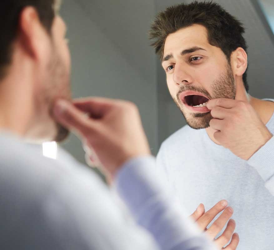
You're likely in the know about common secondary ailments that can impact your dental health. But are you familiar with Eagle syndrome? Many people are not. This syndrome expresses itself as throat and facial pain and is typically associated with the removal of tonsils or trauma to the throat area.
If this sounds like something you could be dealing with, contact your dentist right away. In the meantime, here's what to look out for and what to expect from your care.
Signs of Eagle Syndrome
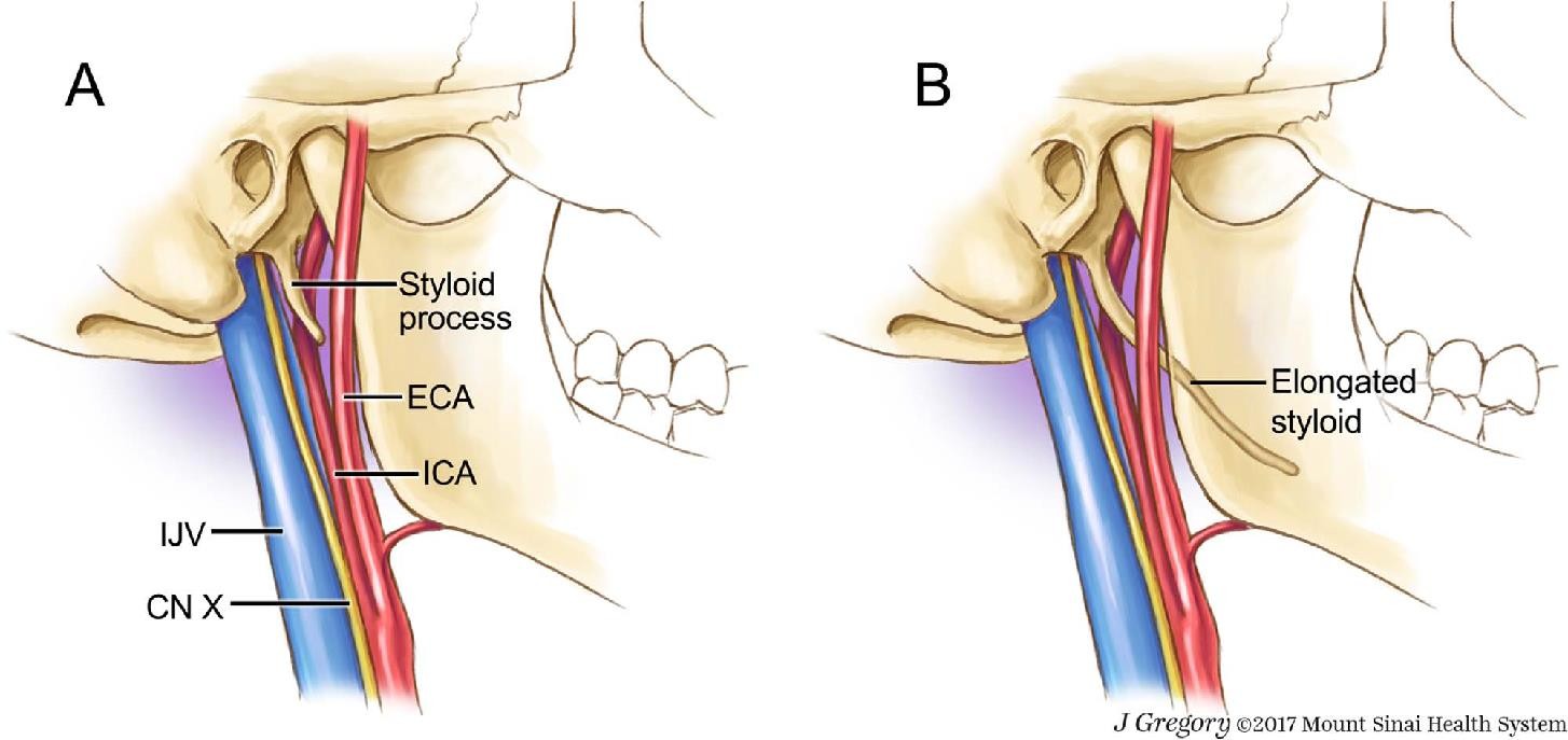
What is Eagle syndrome? Eagle syndrome is also known as an elongated styloid process or styloid-stylohyoid syndrome. The styloid process is a small bone located just below your ear. This small bone can cause a lot of pain if elongation or if calcification occurs. These things result in pinched vessels or nerves and lead to inflammation.
Let's go over Eagle syndrome symptoms so you know what to look for. They include: Sore throat, Earache, Reduced hearing, Tinnitus, Trouble swallowing or chewing, Feeling as though you have something in your throat, Pain when yawning or turning your neck, Facial pain.
According to the Genetic and Rare Diseases Information Center (GARD), only 4 percent of the population have an elongated styloid process, and most patients are asymptomatic. Eagle syndrome is very rare. It's estimated to occur in 1 of 62,500 people, and women are three times more likely than men to have this syndrome.
Diagnosis of Eagle Syndrome
Diagnosis of Eagle syndrome can be challenging because there are many illnesses associated with having a sore throat. Your first response may be to visit your doctor, which is always a good idea. But it's also important to schedule an appointment with your dentist. They can examine your mouth for signs of other problems and recommend the best next steps.
Your doctor or dentist will probably feel your head and neck for any signs of an unusually long styloid process. They may also use an X-ray or CT scan to see your styloid process and stylohyoid ligament in better detail.
You might also be asked to see an ear, nose, and throat specialist to rule out any other conditions that could be causing your symptoms.
Surgical and Non-Surgical Treatments
If you get diagnosed with Eagle syndrome, your medical team will decide the best way to treat it based on your specific case and pain level. Eagle syndrome treatment usually starts with conservative medical management before surgery of any kind is considered.
According to Medscape, medication treatment may include:
Pain relievers, Seizure medication, Antidepressants, Local application of steroids or numbing agents.
Suppose non-surgical, treatment isn't working for you. In that case, your medical team may recommend steroids, pain block injections, or surgery to remove the bone, according to a report published in the Journal of Maxillofacial and Oral Surgery. After surgery, you may receive an analgesics prescription, and your provider will ask you to return in seven days so they can remove your stitches.
You're now informed on the ins and outs of Eagle syndrome. If you're having trouble pinpointing what's happening with throat and facial pain, check-in with your dental professional, they're a fantastic resource to help you with pain in the area. If you're diagnosed with Eagle syndrome, remember that there are plenty of treatment options. And that medicine is usually the first choice before surgery. You've made a great choice to read up on this condition.
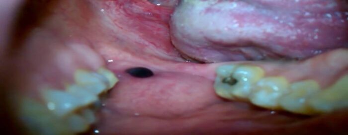
Looking in the mirror and seeing a black spot inside the cheek can be alarming, but is it something serious? The good news is that a dark spot inside your cheek is probably harmless. Consider these possible causes and see your dentist for a definitive diagnosis.
Amalgam Tattoos
If the spot inside your cheek is dark blue, gray or black, it could be a leakage from a dental filling. Amalgam tattoos aren't the permanent ink decorations on your skin that you can have done at a tattoo parlor. Amalgam is the silvery substance that your dentist uses to fill cavities, and it's made from a mixture of tin, zinc, mercury, copper and silver. Sometimes, after a dentist has filled a tooth, the filling mixture leaks, leaving behind a flat, painless dark spot that doesn't grow or change shape.
According to Brigham and Women's Hospital Division of Oral Medicine and Dentistry, amalgam tattoos are permanent, but they don't cause any harm. If the mark is inside your cheek, it's unlikely anyone will see it. However, if you think the spot looks unsightly, speak with your dentist about removal options.
Smoking
Smoking can leave dark stains inside the cheeks and other areas of the mouth, such as on the gums. This condition is called smoker's melanosis, and according to a study in Case Reports in Dentistry, approximately 22 percent of smokers may notice this kind of discoloration in their mouths. It occurs when the tobacco stimulates excessive melanin deposits on the inner lining of the mouth, resulting in a darker pigmentation.
While the condition is benign, patients should keep the other oral effects of tobacco use in mind and consider quitting.
Other Causes of a Black Spot Inside the Cheek
When you see a black spot inside your cheek, you may immediately be concerned that it is cancerous. Rest assured that this is not likely the case, and that treatment may not be necessary.
The Oral Cancer Foundation lists several causes of dark spots inside cheeks that aren't related to cancer. For example, the inner lining of your cheek may just be patchily pigmented. You could have a benign melanotic macule, which is a spot similar to a freckle that can appear in the oral cavity. Alternatively, if you have put pencils in your mouth in the past, the graphite may have become embedded in your mouth lining, creating a dark spot.
Very rarely, a black or dark spot on the inside of the cheek could be a sign of oral malignant melanoma or another type of oral cancer. For this reason, it's always worth seeing your dentist if you notice an abnormal spot in your mouth that doesn't go away, bleeds or grows larger. Your dentist can diagnose the cause of the black spot through an examination, and they may take a sample to send for analysis.
Maintaining a good oral health routine can reduce your fears about a black spot inside your cheek. As well as brushing your teeth twice a day and flossing once a day, regularly check inside your mouth to make sure your gums and the lining of your cheeks look healthy. Report any concerns or symptoms to your dentist, and you can work together to find a solution.
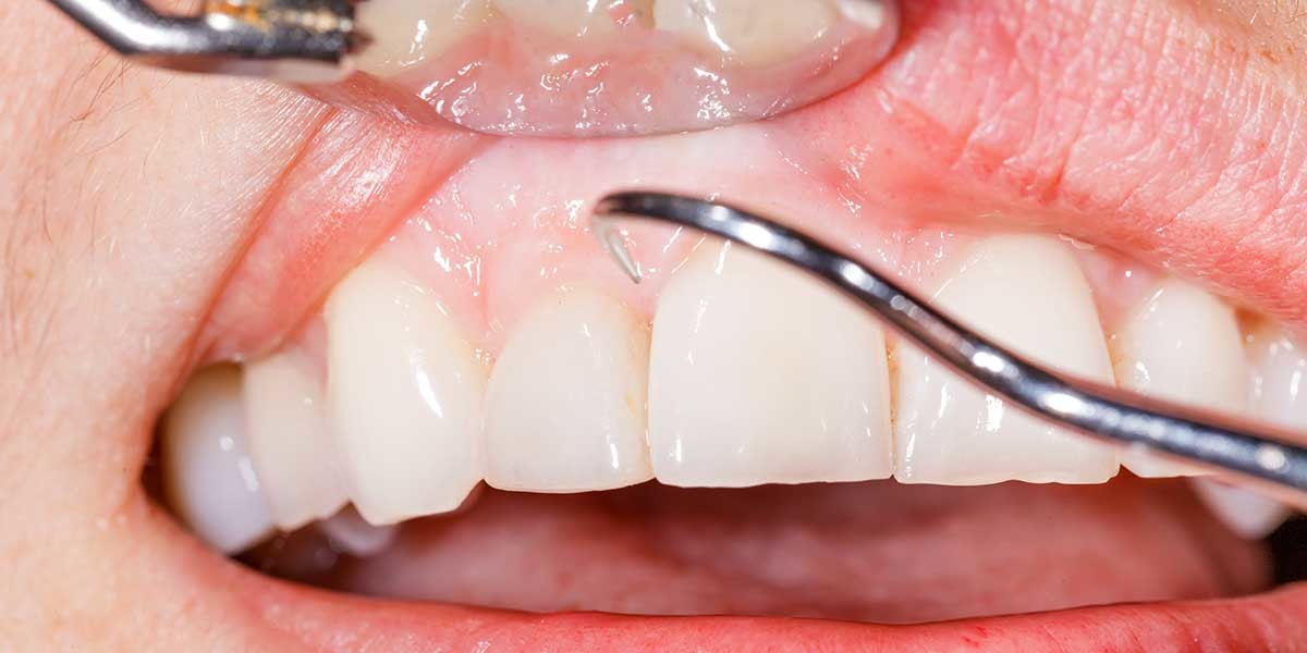
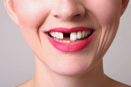
Experiencing your first loose tooth is a right of passage for children. Each primary tooth will fall out and be replaced with a permanent tooth. But as an adult, tooth mobility can definitely cause concern. If you notice that your teeth seem a bit loose, here’s what could be happening.
What Causes Loose Teeth and How Can You Treat It?
Periodontal disease. Untreated gingivitis can advance and become periodontitis, which can eventually cause loose teeth and tooth loss. How? Periodontal disease affects not only the gums but also the tissues and bones that support the teeth. Gums will separate from the teeth, forming pockets between the teeth and gums that become infected. As your periodontitis progresses, the pockets deepen, destroying more gum tissue and bone along the way and eventually causing loose teeth. However, there's some good news: if you pay close attention to your mouth, you can catch the early symptoms of gum disease before it causes loose teeth. And if you do end up with advanced periodontitis, many non-surgical and surgical treatments are available to you, including deep cleaning (scaling and root planing), pocket reduction surgery, soft tissue grafts, and bone grafting.
Pregnancy gingivitis. If you’re pregnant, your mouth can be affected by hormonal changes. Pregnancy gingivitis is an example of this. If you experience inflammation of the gums, or if your gums bleed when you brush or floss, it’s important to consult with your dentist. If left untreated, pregnancy gingivitis can result in periodontitis and, eventually, loose teeth.
Osteoporosis. The part of the jawbone that supports the teeth is known as the alveolar process. An onset of osteoporosis can cause bone loss in the jaw, and studies show a link between a loss of alveolar bone and an increase in tooth mobility.
The National Institutes of Health notes that women with osteoporosis are three times as likely to experience tooth mobility and tooth loss than those who don’t share the disease. You should consult with your dentist or primary care physician if you experience loose teeth. However, if your tooth mobility symptoms are linked to osteoporosis, and you are receiving treatment for it, the ADA recommends that you tell your dentist about any medications you take. Antiresorptive medications can interfere with certain dental treatments and lead to a rare but serious condition called osteonecrosis, which causes loose teeth.
Trauma. The ligaments and tissues that hold your teeth firmly in their sockets can become stretched if you experience an accident or any trauma that extends force to your mouth. This could result in your tooth loosening. For example, loosening can occur from getting hit in your face with a ball. Regularly grinding your teeth can also cause a similar problem. Consider an injury like this to be a dental emergency, and immediately make an appointment with your dentist.
No matter what causes your loose teeth, you’re bound to be worried. But remember, having loose teeth doesn’t mean you have to lose your teeth. As soon as you notice any sign of a tooth or teeth loosening, seek dental care immediately. Your dentist will help you find an effective treatment plan that can save your teeth.
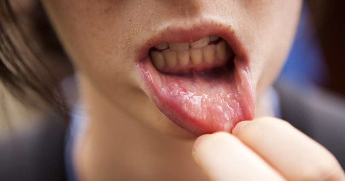
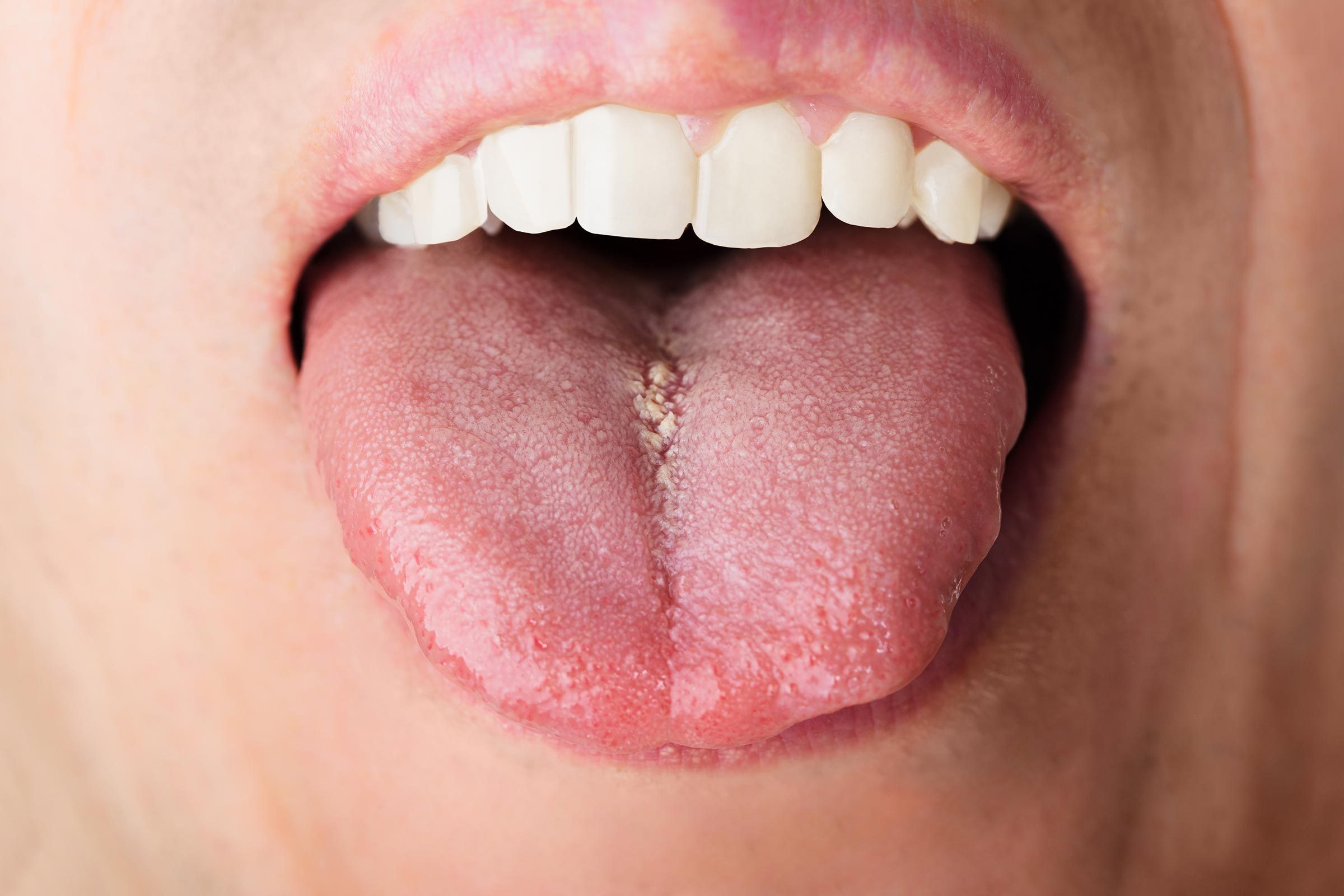
As small as it is, the tongue is one of the strongest muscles in your body, particularly when chewing and swallowing food. And although five-year-olds use it to express their disapproval, it's prone to certain ailments of its own. For example, if you have anemia, tongue function and appearance can feel the effects almost as much as your energy level.
What Is Anemia Tongue
Also referred to as glossitis, explains Healthline, this condition causes the tongue to become inflamed, and is characterized by several things when your iron levels are low. The tongue's appearance can morph into multiple shades of red, and swell slightly in size. The surface of the tongue can smooth out and hide its natural texture, as well. These small bumps you feel on your tongue – also known as papillae – play a crucial role in the eating process, and thousands of taste buds are housed inside them. Papillae alteration can affect how you eat and speak.
Signs and Symptoms
If you think you're suffering from an anemia tongue, schedule an appointment with your dentist so he or she can properly diagnose you. Here are some traits to look for before making the call:
Swollen tongue
Change of tongue color
Difficulty or inability to chew, swallow or speak
Tongue pain and tenderness
Reduction in or loss of tongue papillae
Causes
A variety of conditions can lead to tongue inflammation, some more common than others. According to the National Library of Medicine, these include:
Allergic reactions. Medications, hot or spicy foods and even certain types of mouth care products can irritate the tongue's papillae. Solutions like Colgate® Peroxyl® Mouth Sore Rinse may therefore substitute a less-sensitive mouth rinse.
Injuries. Any sort of mouth trauma resulting from burns or the use of oral appliances like dentures can inflame the tongue.
Oral herpes. Certain diseases, such as oral herpes simplex, can cause blisters, swelling and tongue pain.
Dry mouth. Saliva is a necessity to keep the tongue moist and free of bacteria that can aggravate the tongue's surface.
Of course, the low iron levels defining anemia are your first stop. Iron aids the body in making red blood cells. When you're deficient in them, the tongue's tissue receives a lack of oxygen, much like the rest of the body.
Treatment Options
A trip to your dentist is the best place to start if you suspect you have anemia tongue. During an exam, your dentist will look for blisters, a lack of papillae and any signs of inflammation on your tongue. Blood and saliva samples might also be requested for further testing.
At home, antibiotics, diet changes and proper oral care are all forms of treatment you can use to combat glossitis. Keep in mind a healthy mouth starts with good brushing and flossing. Keep your teeth and gums as healthy as they can be, and being anemic won't mean being in oral pain.
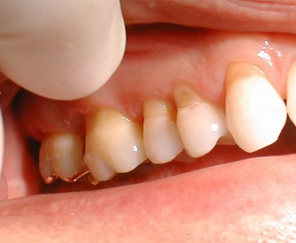
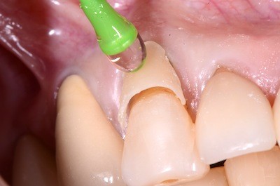
If you've started to notice dents in your teeth where the tooth and the gums come together, you could have abfraction lesions forming. There is no need to worry, though. Your dentist can diagnose the problem and help you find the right treatment plan. Before you go to the dentist, here is what you should know about abfraction lesions.
What Are Abfraction Lesions?
Abfraction lesions are losses of tooth structure. The lesions occur gradually, The lesions occur gradually, with an indentation forming on the front of the tooth near the gumline that gets deeper with time. Abfractions are not cavities but are instead known as non-carious cervical lesions or NCCL. However, because they expose the softer portions of the teeth, like dentin, they can cause tooth sensitivity and mimic the symptoms of a cavity. Discovering the cause is an essential first step to treatment and management.
What Causes Abfraction Lesions?
Abfraction lesions have been attributed to excessive force placed on the teeth during chewing or teeth grinding. However, according to a review in Clinical, Cosmetic, and Investigational Dentistry, there are many factors, including chemical, biological, and behavioral factors, that may contribute to the development of abfractions.
For example, erosion and abrasions can also contribute to the formation of dental abfraction lesions. Tooth erosion occurs from exposure to acids, such as acid reflux or acidic foods and drinks. Tooth abrasion may be caused by improper brushing technique or the use of abrasive toothpastes. This combination causes gum recession and exposes the softer, less mineralized parts of the teeth called the cementum and dentin. Acidic and abrasive factors initiate the abfractions, but often stress from biting can deepen the lesion over time.
How Do You Treat Abfraction Lesions?
Proper abfraction treatment is based on the severity of the lesion and the reported sensitivity and aesthetic concerns. A dentist will usually fill the lesion when it extends below the gums, becomes decayed or challenging to clean, or exposes the tooth's pulp or nerve. Filling the lesion reduces sensitivity and restores the tooth structure. Your dentist may use composite or tooth-colored fillings to cover the notches and improve your smile's appearance.
If teeth grinding causes your abfractions, your dentist may fit you with a mouthguard to protect your teeth while you sleep. Orthodontics can also help prevent further abfraction lesions by realigning your bite and taking pressure off of certain areas of your mouth that may be prone to the damage. Although it won't cure abfractions, try a desensitizing toothpaste if your abfraction is minor. They help relieve the pain associated with tooth sensitivity and work for fast relief.
Your dentist and dental hygienist know how to recognize and modify risk factors for abfractions. If you have tooth sensitivity and you've noticed a lesion starting to form, there is no reason to worry. Talk with your dentist, and they will work with you to decide the best treatment plan for your smile.
A healthy smile involves more than just strong, shiny teeth. Your gums also play an important role in your oral health, and some forms of gum disease can be pretty sneaky. Apical periodontitis, also known as periapical periodontitis, does not always have symptoms but should not be ignored. Find out more about periapical periodontitis and its causes, symptoms, and treatments.
What Is Apical Periodontitis?
Apical periodontitis refers to the inflammation of the periodontium — the tissue that surrounds your teeth. Apical means "relating to the apex," so inflammation usually occurs around the tip — or apex — of the tooth's root. Two types of apical periodontitis exist:
Asymptomatic. Asymptomatic apical periodontitis does not produce any clinical signs or symptoms. However, long-term inflammation can eventually destroy the tissue surrounding the teeth. This type usually develops gradually and is ongoing, which is why it once was referred to as chronic periapical periodontitis.
Symptomatic. Symptomatic apical periodontitis causes pain and discomfort when a person bites down or makes contact with the surrounding teeth. This type of apical periodontitis is usually acute, meaning it comes suddenly and gets worse quickly. However, it can also be chronic.
What Causes Apical Periodontitis?
Typically, apical periodontitis occurs when there's another problem with the tooth. For example, inflammation can develop if a person has an untreated cavity. In some cases, apical periodontitis can develop if the pulp of the tooth becomes infected or dies. Injury or trauma to the tooth can also lead to apical periodontitis.
Because symptoms do not always accompany apical periodontitis, you might not detect it on your own. If you experience pain or discomfort when biting or brushing your teeth, make an appointment to immediately see your dentist. Otherwise, regular dental exams allow your dental professional to note any changes to your oral health and catch asymptomatic inflammation early.
How Do You Treat Apical Periodontitis?
If your dentist notices any inflammation in your gums, they will most likely refer you to an endodontist for treatment. Your treatment depends on what procedures have already been performed and the degree of inflammation. Apical periodontitis treatments could include:
Root canal. In some cases, a root canal can minimize the inflammation of your gums by removing the bacteria and infected tissue from the tooth's pulp.
Apicoectomy. If the infection develops or continues after the root canal, you might require an apicoectomy. During this procedure, the endodontist removes the tip of the tooth's root and infected tissue, then seals the end of the root with a filling.
A proper oral hygiene routine and regular visits to the dentist can help protect your gums from apical periodontitis. If you notice any pain or swelling in your gums, schedule an appointment with your dentist. They can take a look inside your mouth, assess your symptoms, and recommend the appropriate next steps.