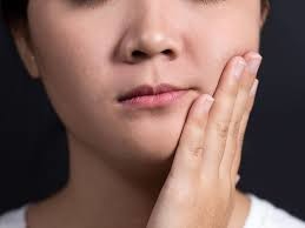
While we wish our teeth were as strong as metal, they’re susceptible to wear and tear—and sometimes that can come in the form of a crack. There are five types of cracked teeth, and the one we’ll discuss today is called a vertical root fracture. We’ll look into the fractured tooth root symptoms, causes, and treatment.
Imagine looking at a whole tooth and seeing a line at the very bottom moving upwards. That’s what a vertical root fracture looks like. Vertical root fractures are vertical cracks in your tooth that begin at the root of your tooth and run toward the top of the tooth. Because they normally don’t show signs or symptoms, they can go unnoticed. However, they can lead to an infection in the surrounding bone and gum, which can be noticeable and painful for the patient.
Why Do Vertical Root Fractures Happen?
These fractures are commonly found in weak teeth, teeth that have been treated with a root canal, and other restorative treatments like crown placements. Healthy teeth can also be subject to cracks by chewing on hard materials and foods, like ice. Specifically, when a root canal is being performed, a jolt of pain, sound of popping, or bleeding in the canal might also be a sign of a vertical root fracture that happened during the procedure.
Symptoms of a Vertical Root Fracture
Symptoms of fractures can vary, making this a difficult condition for dental professionals to diagnose. However, signs that you may have a vertical root fracture include:
Mild pain when biting
The appearance of a crack when examining with a special light or dye
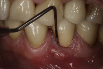
A draining sinus tract appearing next to the tooth that looks like a boil or ulcer, which is often a sign that there is an infection beneath the tooth
A pocket between the gum and the tooth, near the fracture, where the gum essentially detaches from the tooth
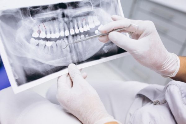
A dental professional or specialist (usually an endodontist) may diagnose a fracture during a root canal procedure if they can see a crack. If the fracture occurs after the procedure, they might need to take X-rays, which may show the fractured root characteristically shaped like the letter J, or the specialist may also use a diagnostic method known as transillumination, where they shine a light through your tooth to detect any fracture lines.
How Are Vertical Root Fractures Treated?
There are a few different treatments for a tooth that has a vertical root fracture. In some cases, special types of cement are used to bond the teeth or stop the propagation of the fracture. In most cases, however, the most common treatment is tooth extraction. It's important to have this procedure as soon as a fracture is diagnosed, as chronic infection can eat away at the bone around the tooth, which may be needed in the future to support a dental implant.
To avoid dealing with the fallout of a vertical root fracture, here are some preventive steps you can take to protect your teeth:
As these fractures occur mostly in teeth treated with a root canal, avoid this treatment if possible. This means scheduling regular dental examinations so your dentist can detect problems early and help you prevent cavities from forming in the first place.
If you need root canal treatment, ask your endodontist about the root canal filling technique they will use and discuss how to reduce your fracture risk, such as avoiding post-placement.
If your symptoms match those outlined above and you're worried that you may have this type of fracture, talk with your dental professional. They will help you determine the cause of your symptoms and give you the proper treatment to keep your teeth healthy and functioning—so you can worry less and smile more.
A fistula is a canal that develops between two points to drain an infection from an abscess, and a sinus tract is a drainage canal that originates at a point of infection but has only one ending. Although these terms are used interchangeably in relation to dentistry, a dental fistula is more likely to be a sinus tract infection than an actual fistula. A fistula or tract can take various forms, and the cause and location of the tract helps determine the best treatment method.
Causes
A dental or oral fistula is commonly associated with an abscess, which can be caused by trauma or injury to the mouth, a build-up of food bacteria, or be the result of surgery, extraction of the molars, or a congenital defect. Whether the abscess is gum- or tooth-related, it usually results in an infection that can spread to the bone or the tooth pulp. As the pressure and pain of the abscess builds, the infection pushes its way to the surface of the gum to drain. Occasionally, a failed root canal treatment might cause a pus corridor to form near the location of the root tip or apex.
Symptoms
The typical indication of a dental fistula or sinus tract is a bump that develops on the gum tissue or gingiva, where it’s called a gum boil, or in proximity to an abscessed tooth. The bump might alternatively appear and disappear, and is a sign that infection exists and your body is using the fistula to drain it. Since draining releases the pressure of the abscess, the fistula itself is often not painful, although many patients report an unpleasant taste.
Treatment
Since an infection is the root cause, the fistula is unlikely to heal and disappear on its own. Without care, the infection can travel to your jawbone and affect other parts of the body. Here are a few treatment options your doctor may prescribe:
Rinse with a warm salt water solution to kill bacteria.
Schedule an urgent dental examination to determine the severity of the infection.
Take antibiotic medication prescribed by your doctor or dentist, to help contain the infection.
Maintain your oral hygiene regimen to ensure the healthiest environment possible.
Your dental professional’s first line of defense will be to clean the area around the fistula, and allow accumulated pus to run out. Most dentists recommend antimicrobial therapy to help your body fight the infection. For bacterial infections that begin inside a tooth, the dentist will make a small hole in the tooth enamel to enable the infection to drain. In the case of a badly infected and damaged tooth, your dentist may elect to extract the tooth. This process may be followed by a root canal treatment or an apicoectomy to remove the tip, and a discussion of your options for replacement of the tooth.
Prevention
The prognosis for a dental fistula is typically very positive, provided you follow recommended treatment to eliminate the infection. Prevent future infections keeping up your daily brushing and flossing routine, supplemented by regular dental exams and cleanings.
Hydrated Silica might sound like a complex scientific name, hydrated silica is simply a hydrated form of silicon dioxide. As the Encyclopedia Britannica explains, silica is the second-most abundant mineral in the Earth's crust. It's naturally crystalline, with sand and obsidian being common forms.
To make hydrated silica, the silica crystals must be heated and dissolved in water to create liquid sodium silicate. The liquid form is then mixed with acid and precipitated, which turns it into a solid. The end result is a fine, white powder, or granules. Depending on the size of the particles, they can then be used in a variety of ways.
What Does Hydrated Silica in Toothpaste Do?
Depending on the specific formulation and particle size, hydrated silica can have several uses in both natural and conventional toothpaste. According to an article in the Journal of the Pakistan Dental Association, hydrated silica acts as an abrasive to help to remove plaque and other particles from your teeth when brushing with toothpaste. Besides aiding in the removal of plaque and food particles, abrasives like hydrated silica can also help to remove stains, which makes them useful as whitening agents in toothpastes as well. Abrasives used in toothpaste date back over 2000 years, where paste mixtures were once made with bones and ground shells.
In addition to removing particles and minimizing stains on your teeth, hydrated silica can also act as a thickening agent to help form the toothpaste's consistency.
Is Hydrated Silica Safe?
While the word "abrasive" may sound scary, these ingredients are designed to help scrub away plaque and stains without harming your teeth. Hydrated silica is an ingredient that has been in use for a long time and which the Food and Drug Administration (FDA) lists as Generally Recognized As Safe (GRAS). According to the FDA's Select Committee on GRAS Substances, silicates are biologically inert, which means they don't create a reaction in the body, and there is no evidence to suggest that they could pose a hazard.
Not only is hydrated silica safe to use every day in oral care products, but you can also feel confident choosing products with this ingredient because it is derived from one of the most abundant natural materials on the planet, making it a renewable natural source. The Environmental Working Group also gives hydrated silica a score of one out of ten, which means that it poses the lowest possible hazard to people and the environment based on their data.
It's not always easy to take time out of your day to research ingredients, but you don't need to earn a science degree to understand what's inside your toothpaste. I did just a little bit of reading—and I'm glad I did, because I learned about the many uses and safety of specific ingredients, such as hydrated silica. Plus, I finally understood where they actually came from. I then felt more equipped to make a confident and informed choice when I made the switch. After all, no matter how carefully you read that ingredient list, it's hard to find any answers without knowing what the words mean.
When you see hydrated silica listed on your oral care products, you can feel confident that you understand what its function is, how its derived, and why it's safe to use.
Diarrhea and cramps are among the most well-known ulcerative colitis (UC) symptoms. They stem from inflammation and sores called ulcers in the intestines. What you may not realize is ulcerative colitis sores can form in any part of your gastrointestinal (GI) tract, from your mouth to your anus.
Mouth problems can start even before more typical symptoms like cramps and diarrhea. Some mouth sores are short-lived and are more of a nuisance than a real problem. Others can affect your ability to talk or eat and require your doctor’s help to manage.
Symptoms of mouth problems
Ulcerative colitis mouth ulcers are often associated with the following symptoms: Pus-filled sores, canker sores, dry mouth, mouth pain, swollen tongue, bad breath, metallic taste or other unusual taste in the mouth.
Causes
Swelling and ulcerative colitis sores can appear anywhere in your GI tract, including in your mouth. Ulcerative colitis mouth ulcers can also be a side effect of some ulcerative colitis medications that cause dry mouth and swelling in the mucous membranes.
Vitamin and mineral deficiencies can also lead to ulcerative colitis mouth sores and other problems. Inflammation in your intestines can make it harder for your body to absorb nutrients like B vitamins and iron from food. You can also lose these nutrients when you have diarrhea.
Common mouth problems
Ulcerative colitis and its treatments can cause the following mouth problems: Mouth sores, Dry mouth, Bad breath, Taste changes, Inflamed lips.
Treatment
The first step to relieving ulcerative colitis mouth sores and other mouth problems is to reduce inflammation in your gastrointestinal tract and get your ulcerative colitis under control.
Medications like aminosalicylates (5-ASAs), corticosteroids, immunomodulators, and biologics calm the overactive immune system response that causes inflammation and sores. Your doctor will help you find the right drug or drugs to manage your ulcerative colitis.
An antiseptic mouthwash can help keep your mouth clean while your sores heal. Taking a multivitamin or mineral supplement and eating a balanced diet helps to prevent the nutrient deficiencies that can cause ulcerative colitis mouth ulcers and other mouth problems.
Talk to your doctor if you think a medication you take for ulcerative colitis could be causing these symptoms. Your doctor may recommend alternative treatments less likely to cause mouth sores or suggest other ways to manage this side effect.
When to see a doctor
Let your doctor know if you have any new symptoms in your mouth or other parts of your digestive tract. Also call if your mouth problems affect your ability to eat or talk.
The takeaway
Mouth problems aren’t the most common symptoms of ulcerative colitis. They sometimes appear before more common symptoms such as diarrhea and stomach cramps. Watch for sores, swelling, pain, and taste changes and report them to your doctor. Changing your treatment or adding a nutritional supplement may help to relieve these issues.
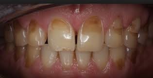
Enamel is the thin outer covering of the tooth. This tough shell is the hardest tissue in the human body. Enamel covers the crown which is the part of the tooth that's visible outside of the gums.
Because enamel is translucent, you can see light through it. But the main portion of the tooth, the dentin, is the part that's responsible for your tooth color -- whether white, off white, grey, or yellowish.
Sometimes coffee, tea, cola, red wine, fruit juices, and cigarettes stain the enamel on your teeth. Regular visits to your dentist for routine cleaning and polishing can help remove most surface stains and make sure your teeth stay healthy.
What does tooth enamel do?
Enamel helps protect your teeth from daily use such as chewing, biting, crunching, and grinding. Although enamel is a hard protector of teeth, it can chip and crack. Enamel also insulates the teeth from potentially painful temperatures and chemicals.
Unlike a broken bone that can be repaired by the body, once a tooth chips or breaks, the damage is done forever. Because enamel has no living cells, the body cannot repair chipped or cracked enamel.metimes coffee, tea, cola, red wine, fruit juices, and cigarettes stain the enamel on your teeth. Regular visits to your dentist for routine cleaning and polishing can help remove most surface stains and make sure your teeth stay healthy.
What are the environmental causes of tooth surface erosion?
Friction, wear and tear, stress, and corrosion (or any combination of these actions) can cause erosion of the tooth surface. More clinical terms used to describe these mechanisms include:
Attrition. This is natural tooth-to-tooth friction that happens when you clench or grind your teeth such as with bruxism, which often occurs involuntarily during sleep.
Abrasion. This is physical wear and tear of the tooth surface that happens with brushing teeth too hard, improper flossing, biting on hard objects (such as fingernails, bottle caps, or pens), or chewing tobacco.
Abfraction. This occurs from stress fractures in the tooth such as cracks from flexing or bending of the tooth.
Corrosion. This occurs chemically when acidic content hits the tooth surface such as with certain medications like aspirin or vitamin C tablets, highly acidic foods, GERD, and frequent vomiting from bulimia or alcoholism.
What are the signs of enamel erosion?
The signs of enamel erosion can vary, depending on the stage. Some signs may include:
Sensitivity. Certain foods (sweets) and temperatures of foods (hot or cold) may cause a twinge of pain in the early stage of enamel erosion.
Discoloration. As the enamel erodes and more dentin is exposed, the teeth may appear yellow.
Cracks and chips. The edges of teeth become more rough, irregular, and jagged as enamel erodes.
Severe, painful sensitivity. In later stages of enamel erosion, teeth become extremely sensitive to temperatures and sweets. You may feel a painful jolt that takes your breath away.
Cupping. Indentations appear on the surface of the teeth.
When enamel erodes, the tooth is more susceptible to cavities or tooth decay. When the tooth decay enters the hard enamel, it has entry to the main body of the tooth.
ow is tooth enamel loss treated?
Treatment of tooth enamel loss depends on the problem. Sometimes tooth bonding is used to protect the tooth and increase cosmetic appearance.
If the enamel loss is significant, the dentist may recommend covering the tooth with a crown or veneer. The crown may protect the tooth from further decay.
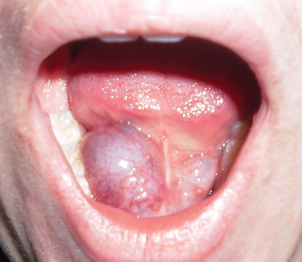
A ranula is a fluid collection or cyst that forms in the mouth under the tongue. It is filled with saliva (spit) that has leaked out of a damaged salivary gland. Salivary glands are small structures around the mouth which make saliva. Saliva should drain from these glands directly into the mouth. If one of these glands is damaged then the saliva leaks out into the tissues next to the gland forming a cyst or bubble near the gland. This cyst is called a ranula.
If the ranula stays in the mouth underneath the tongue it is called a simple ranula and if it grows down into the neck under the mouth it is called a plunging ranula.
How are Ranulas Diagnosed?
The diagnosis is usually easy for an experienced physician to make based primarily on physical examination but often specialized radiology studies such as Ultrasound, Computed Tomography (CT Scan), or Magnetic Resonance Imaging (MRI) are needed to define the full extent of the problem and exclude other causes for swelling. No other laboratory tests are usually needed to make the diagnosis.
What are the Symptoms of a Ranula?
Ranulas are usually discovered by the patient, the patient’s family, or the patient’s medical caregivers like medical doctors and dentists. It usually is a 2-3 inch diameter painless soft swelling under the tongue or chin that is easy to identify. Occasionally, the fluid collection can hurt a little and sometimes is can spontaneously empty into the mouth only to slowly fill back up in the weeks after it empties. Usually, it just slowly grows in size until it is discovered.
Rarely, a ranula can spontaneously go away without any treatment but usually a procedure will be needed to treat the problem. Simple drainage of the fluid collection rarely permanently fixes the problem as the diseased gland continues to leak saliva.
What are the Potential Complications of This Treatment?
Our percutaneous treatment is safe and effective. The only common side effect of treatment is mild painless swelling in the treated area for several days after treatment. Patients can eat normally immediately after the procedure and as there are no incisions or stitches, no wound care or dressings need to be managed.
Sometimes, the ranula is not completely cured with one treatment and a second treatment is necessary. Rarely, the diseased gland resists the medication injections and continues to leak saliva necessitating referral to an experienced surgeon for traditional surgical removal.
While our percutanous procedure is very safe, all treatments carry risks. The only serious risk that can occur with alcohol injection under the mouth or chin is injury to a nearby nerve which can result in temporary muscle weakness in the area in rare cases (2-3%).
What is the Outlook for Patients With a Ranula?
Ranulas are benign fluid collections near the mouth which can be effectively treated with our percutanous treatment but also with traditional surgical approaches. Both appraches offer permanent removal of the ranula and resolution of symptoms. We feel our percutanous approach offers a less invasive treatment for this benign condition.
When Should I See a Doctor?
If a ranula is suspected as there is a 2-3 inch diameter soft swelling under the tongue or chin medical attention with the child’s medical provider should be obtained. If the diagnosis is felt to be a ranula based upon their examination and imaging tests treatment can then be sought from specialists such as interventional radiologists or surgeons.
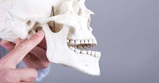
Oral health isn’t just about your teeth. The jawbone is a crucial player in many of your day-to-day functions, such as eating, breathing and speaking.
Since the jawbone is so important, you want to watch out for jaw pain, which could be a symptom of a fracture or dislocation. If you suspect a serious injury, address it as quickly as possible to prevent further jaw damage.
Fractured Jaw Symptoms
If you have recently experienced facial trauma, such as physical assault, a sports injury or a car accident, and you are experiencing jaw pain, your jaw may be fractured. You can test the theory by attempting to open and close your jaw. If something feels off when you do this, or you have lost teeth, this increases the likelihood of a fractured jaw. Assess your situation further by:
Examining your face: Check for swelling, bruising or protrusions on the side of your face.
Evaluating your pain level: Pay attention to the pain in your jaw as you chew and note any increases in your pain level.
If you experience any of these symptoms, seek medical attention immediately. Failure to treat a fractured jaw can result in infection.1
Dislocated Jaw Symptoms
While the symptoms of a dislocated jaw are different than those of a fractured jaw, the consequences of leaving it untreated are much the same. Pain is a symptom as well as: Difficulty speaking, Abnormal bite, An overbite that didn’t previously exist
Treatment Options
While common (only the nose is broken more frequently than the jaw), jaw injuries are treated as emergencies. As you await medical treatment, support your lower jaw to help stabilize it and keep your airway open. For both types of jaw injuries, you will need to see a doctor. Do not attempt to fix your own jaw as this could cause further pain and damage.
A doctor or oral surgeon will manipulate a dislocated jaw into the correct position. They may be able to do this manually after you’ve received anesthetics and muscle relaxants. These medications will minimize pain and allow the doctor to more easily manipulate the jaw.
Depending on the extent of the break, treatment for a jaw fracture may require surgery. Clean breaks may heal on their own while your jaw is immobilized, while multiple fractures will likely require surgical repair.
Recovery
Both dislocated and fractured jaws are bandaged or wired shut during recovery to prevent you from opening your jaw too wide and to keep your bite in its proper place. After resetting a dislocated jaw, your doctor may wrap a bandage around your head and under your chin.
During your recovery, you won’t be able to open your jaw very wide for at least six weeks. Your diet during this time will consist of mostly liquids as you likely won’t be able to chew solid food. A few of the soft foods you may be able to chew depending on your situation include:
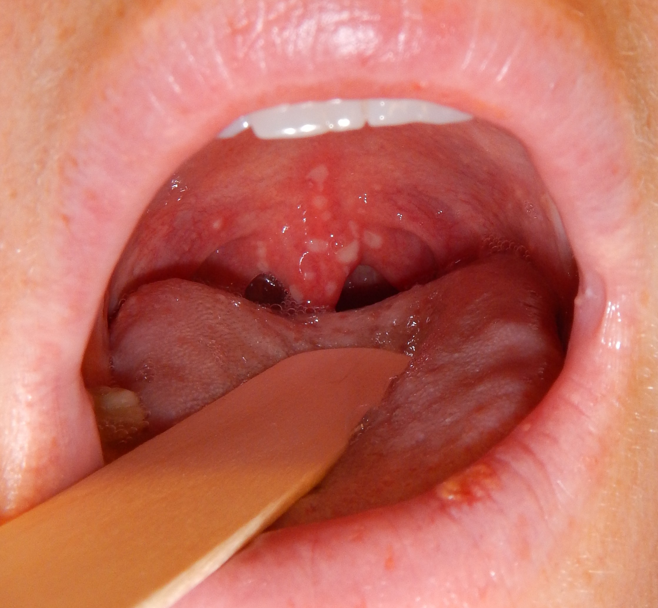
Herpangina is an acute viral illness in children. Common symptoms are small blister-like bumps or sores (ulcers) in the mouth and fever. It is caused by a virus. The most common ones are coxsackie viruses A and B.
What are the symptoms of herpangina?
Symptoms of herpangina typically show up two to five days after you’ve been exposed to the virus. The symptoms of herpangina vary from person to person, but can include: Sudden onset of fever, sore throat, headache, neck pain, swollen lymph glands, difficulty in swallowing, loss of appetite, drooling (in infants), vomiting (in infants).
Small ulcers in the back of the mouth and throat begin to appear about two days after the initial infection. They tend to be light gray and often have a red border. The ulcers usually heal within seven days.
What causes herpangina?
Herpangina is usually caused by group A coxsackieviruses. However, it can also be caused by group B coxsackieviruses, enterovirus 71, and echovirus. Infections caused by these viruses are highly contagious. The viruses can be shared easily between one child and another. They’re most commonly spread through droplets from sneezes or coughs or contact with fecal matter. Proper handwashing can help reduce the risk of sharing the viruses. After a child gets herpangina, they usually develop a natural immunity to the virus that caused it. However, they may still be affected by other viral strains that can cause the illness.
How is herpangina treated?
The primary goal of treatment is to reduce and manage symptoms, especially the pain. Your specific treatment plan will depend on a variety of factors, including your age, symptoms, and tolerance for certain medications. Since herpangina is a viral infection, antibiotics aren’t an effective form of treatment. Antivirals for herpangina do not exist. Instead, your doctor may recommend:
Ibuprofen or acetaminophen
These medications can ease any discomfort and reduce fever. Do not use aspirin to treat symptoms of a viral infection in children or teenagers. This has been linked to Reye’s syndrome, a life-threatening illness that results in sudden swelling and inflammation in the liver and brain.
Topical anesthetics
Certain anesthetics, such as lidocaine, can provide relief for a sore throat and any other mouth pain associated with herpangina. With treatment, symptoms should disappear within seven days with no lasting effects. If the symptoms worsen or linger longer than 10 days, you should see your doctor again.
How can herpangina be prevented?
Practicing good hygiene is the best way to prevent herpangina. You should always wash your hands thoroughly, especially before meals and after using the restroom. It’s also important to cover your mouth and nose when sneezing or coughing to prevent the spread of germs. Teach your children to do the same. While caring for a child with herpangina, wash your hands frequently, especially after coming in contact with soiled diapers or mucus.
Clean any surfaces, toys, and other objects with a disinfectant to kill germs. You should also keep your child out of school or daycare for a few days to avoid spreading the infection to others.
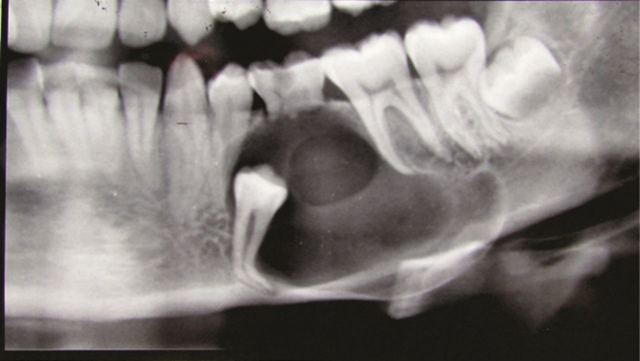
Dentigerous cysts are the second most common type of odontogenic cyst, which is a fluid-filled sac that develops in the jaw bone and soft tissue. They form over the top of an unerupted tooth, or partially erupted tooth, usually one of your molars or canines. While dentigerous cysts are benign, they can lead to complications, such as infection, if left untreated.
What are the symptoms?
Smaller dentigerous cysts might not cause any symptoms. However, if the cyst grows larger than 2 centimeters in diameter, you may notice:
swelling
tooth sensitivity
tooth displacement
If you look inside your mouth, you may also notice a small bump. If the cyst causes tooth displacement, you might also see gaps slowly forming between your teeth.
What causes it?
Dentigerous cysts are caused by a buildup of fluid over the top of an unerupted tooth. The exact cause of this buildup is unknown.
While anyone can develop a dentigerous cyst, they’re more commonTrusted Source in people who are in their 20s or 30s.
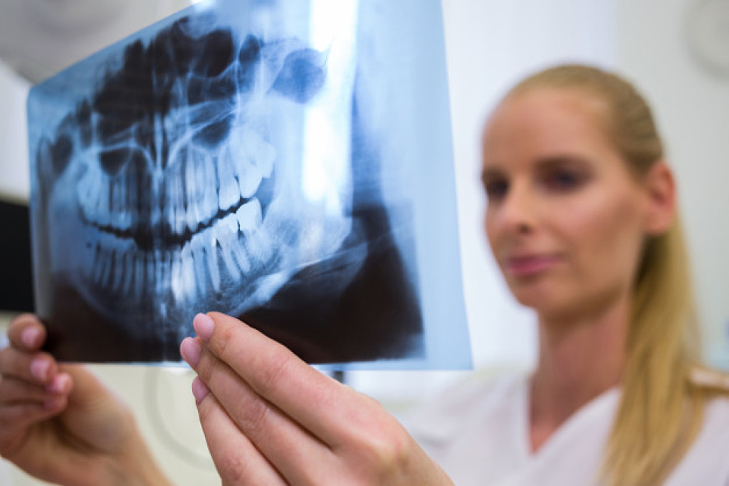
How is it diagnosed?
Small dentigerous cysts often go unnoticed until you have a dental X-ray. If your dentist notices an unusual spot on your dental X-ray, they may use a CT scan or MRI scan to make sure it’s not another type of cyst, such as a periapical cyst or an aneurysmal bone cyst.
In some cases, including when the cyst is larger, your dentist may be able to diagnose a dentigerous cyst just by looking at it.
How is it treated?
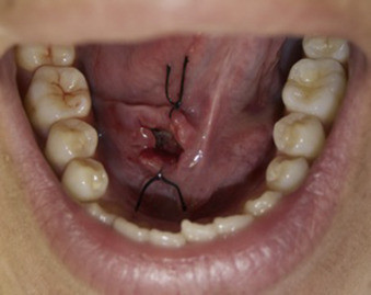
Treating a dentigerous cyst depends on its size. If it’s small, your dentist might be able to surgically remove it along with the affected tooth. In other cases, they might use a technique called marsupialization.
Marsupialization involves cutting open the cyst so it can drain. Once the fluid has drained, stitches are added to the edges of the incision to keep it open, which prevents another cyst from growing there.
What are the complications?
Even if your dentigerous cyst is small and not causing any symptoms, it’s important to have it removed to avoid complications. An untreated dentigerous cyst can eventually cause:
infection
tooth loss
jaw fracture
ameloblastoma, a type of benign jaw tumor.
Living with a dentigerous cyst
While dentigerous cysts are usually harmless, they can lead to several problems if left untreated. Talk to your dentist about any swelling, pain, or unusual bumps in your mouth, especially around your molars and canines. In most cases, dentigerous cysts are easy to treat, either through excision or marsupialization.
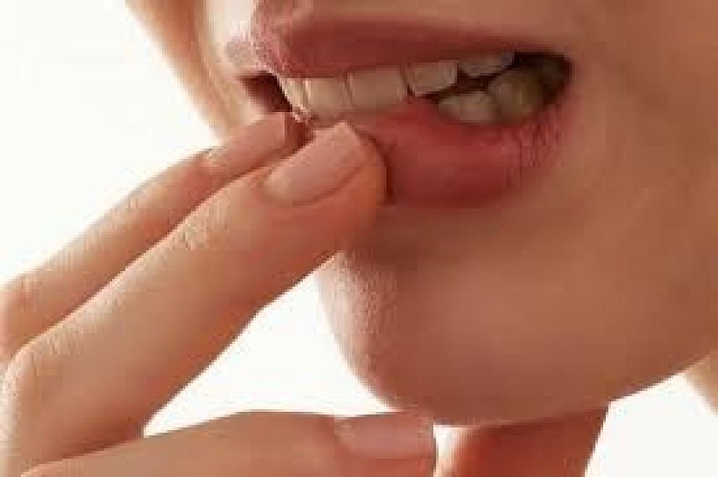
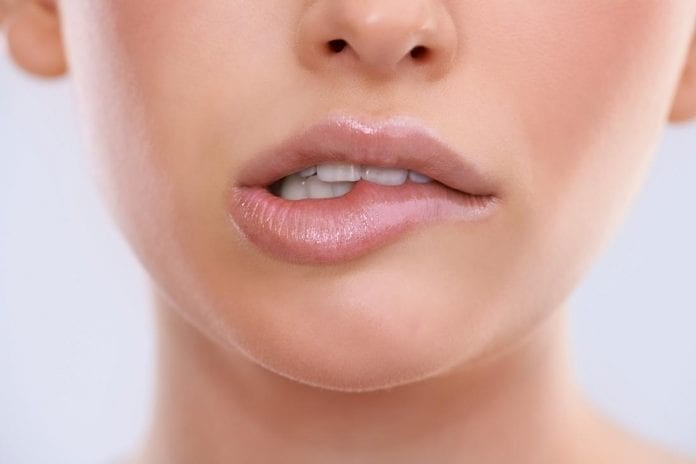
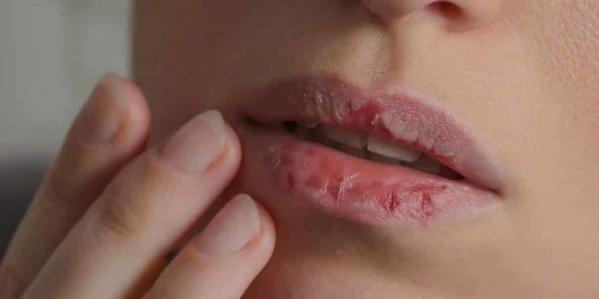
For many people, lip biting is just an occasional nervous habit. However, other people may chronically bite their lips due to an underlying medical condition. Here are some of the many conditions that can cause this oral habit, including dental conditions, psychological conditions and developmental disorders.
Temporomandibular Disorders
The temporomandibular joint (TMJ), which connects your jawbone to your skull, is one of the most complex joints in the body. Disorders of this joint can be caused by numerous factors, such as arthritis or injuries to the jaw. People with TMJ disorders may experience many different symptoms, such as pain in the jaw joint or trouble opening and closing the mouth.
A study published found that lip biting is also a common symptom of TMJ disorders, with 37 percent of the studied individuals exhibiting the habit of biting their lips or other objects.
Malocclusion
Malocclusion means the upper and lower teeth aren’t aligned properly. Teeth may be misaligned if your upper and lower jaws aren’t the same size. Extra teeth, abnormally shaped teeth or missing teeth are some other possible causes of misalignment.
Most teeth alignment problems are minor and don’t need any treatment, although in some cases, individuals may have trouble or discomfort when biting or chewing. Repetitive lip biting in children with an existing malocclusion can impede correction of the improper alignment.
Other Health Conditions
Dental conditions such as a TMJ disorder and malocclusion aren’t the only possible scenarios in which individuals bite their lips. Many other health conditions can also result in this oral habit.
Sometimes, psychological conditions can cause lip biting. Body-focused repetitive behaviors (BFRBs) are one of these disorders. People with BFRBs may repeatedly pull their hair, pick their skin, bite their lips or perform other repetitive actions.
Individuals with autism may also have a tendency toward certain self-harm behaviors, such as biting their lips.
Treatment Options
If you often bite your lips, or if you have a child with this habit, see a dentist. There are many treatments available based on the underlying cause.
If your dentist suspects a TMJ disorder is to blame, they may suggest home remedies such as massaging the jaw muscles or limiting your diet to soft foods. If necessary, they may prescribe medications to help ease pain and inflammation in the jaw joint. Your dentist may even recommend a nightguard or splint, which is a clear plastic device that fits over your teeth, to help your jaw muscles relax. Referrals to other medical specialists, such as physiotherapists or oral surgeons, may sometimes be needed to treat TMJ disorders.Orthodontic treatment may be required to correct malocclusion and its associated issues.
Your dentist may recommend braces or other orthodontic appliances to adjust the positioning of your teeth. If overcrowding is part of the malocclusion problem, one or more teeth may need to be extracted to make room. In rare cases, a patient may need surgery to reshape the jaw.